|
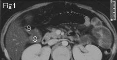
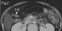
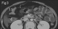
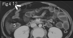
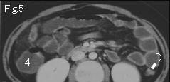
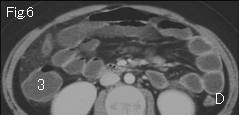
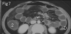
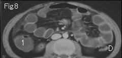
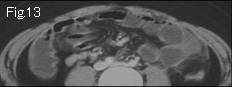
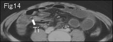
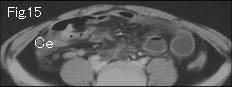
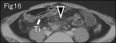
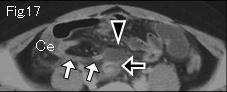
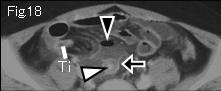
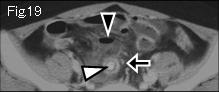
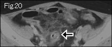
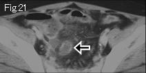
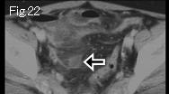
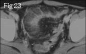
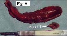
Fig.9-Fig.12 are abbreviated. Small bowel is distended (more than 2.5cm outside diameter) in Fig.1-Fig.20. In Fig.5-Fig.8, ascending colon includes liquid contents same as small bowel, and descending colon (D) is collapsed. On tracking down from ascending colon 1 of Fig.8 to an caudal side to search whether there is an obstructive lesion, 10 of Fig.2 shows collapsed transverse colon. Because transition zone (from 10 of Fig.2 to 12 of Fig.4) does not depict neoplastic lesion, intussusception or compression from outside lesions, distended or fluid-filled intestine indicates paralytic ileus. White arrows of Fig.17 is collapsed proximal appendix and black arrows of Fig.18-Fig.22 is distended appendix which contains fecalith (white arrowhead). Black arrowheads of Fig.16-Fig.19 present extraluminal liquid accumulation and free air, and surrounding fat stranding indicate perforated appendicitis. Ce: cecum, Ti: terminal ileum. The findings were confirmed at surgery (Fig. A: necrotic appendix).
|