|
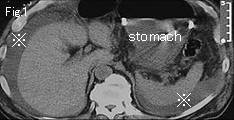
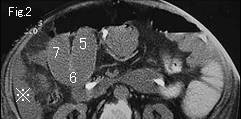
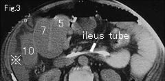
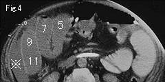
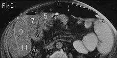
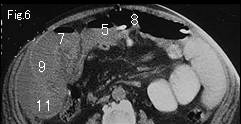
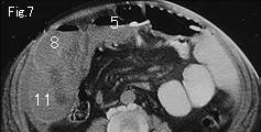
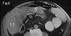
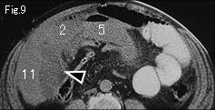
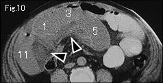
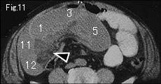
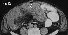
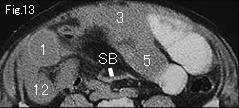
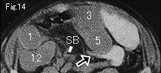
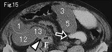
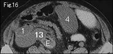
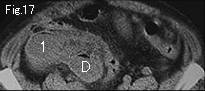
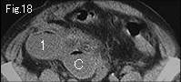
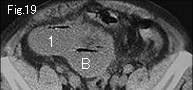
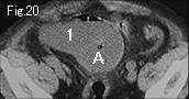
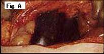
Fig.1-Fig.4 show massive ascites (reference mark) around liver, spleen and in right flank, and Fig.9-Fig.11 show marked mesenteric stranding (black arrowheads). Small bowel contrasted with gastrografin indicates simple obstruction, and non-contrasted bowel is easily recognized as strangulated small bowel. Closed loop formation can be proved by tracing noncontrasted bowel. A of Fig.20 occludes at F of Fig.15, and 1 at 13 (same number as long as goes up and down) of same figure too. A beak sign (black arrows) of simple obstruction site is depicted in Fig.14 and Fig.15, and collapsed small bowel (SB) in Fig.13 and Fig.14 indicating closed loop formation. Necrotic small bowel of 150cm length (Fig. A) was strangulated by an adhesive band formed between sigmoid colon and appendectomy site.
|