|
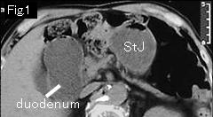
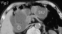
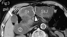
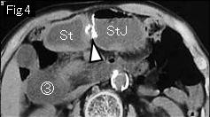
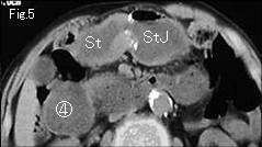
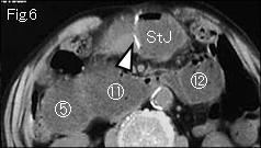
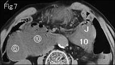
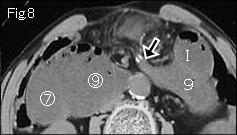
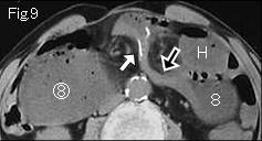
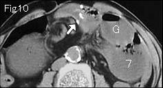
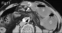
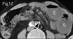
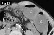
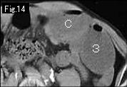
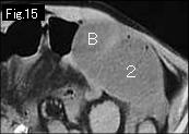
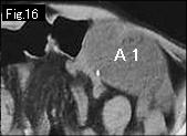
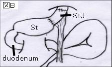
In Fig.1 - Fig.6 "St" is an original stomach, "StJ" is interposed jejunum, and white arrowhead is the anastomosis where staplers are shown. Duodenum is distended on the right side (circle number 1 of Fig.2- circle number 11 of Fig.6 ). There is no ascites, but because of distended gasless small intestine in a left side and mesenteric stranding in Fig.11 and Fig.12.(black arrowhead), strangulated obstruction is still possible. On tracking down from A and 1 of Fig.16 in retrograde fasion , each connects to J and 10 of Fig.7. On the other hand, distended duodenum (circle number 1 of Fig.2) can be traced to jejunum of circle number 12 of FIg.6. The difference of density of fluid content between jejunum (circle number 12 of FIg.6) and J/10 of Fig.7 suggests that circle number 12 of FIg.6 is starting point of simple obstruction, and J/10 of Fig.7 present obstruction site of closed loop. It will be easier to make the diagnosis with enhanced CT scan with difference of mural enhancement effect. Black arrow of Fig.8 and Fig.9 show collapsed distal jejunum. White arrows of Fig.8 and Fig.9 are staplers of anastomosis. Surgery revealed strangulated jejunal obstruction by mesentery of jejunum anatomized in stomach (Fig. B) with no necrosis.
|