CT of Acute Abdomen

Epigastric Pain series 4 RESIDENT COURSE Answer [ER Case 17]

Small bowel necrosis


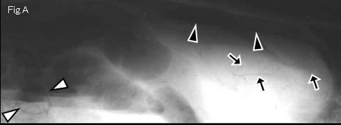
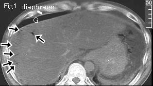
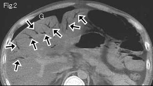
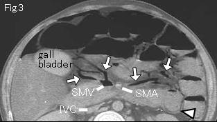
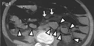
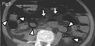
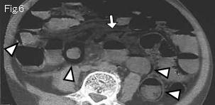
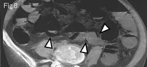
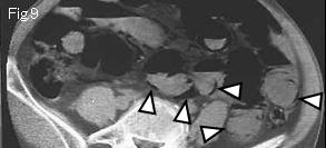
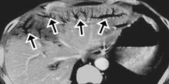
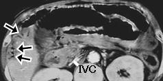
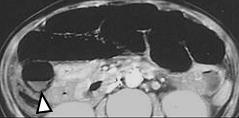
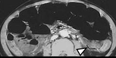
There are three findings to suggest bowel necrosis in abdominal plain film Fig.A. 1)free air (black arrowhead), 2)gas in portal vein (black arrow) which spreads to periphery nearby the liver surface, 3)intramural pneumatosis (white arrowhead) of small bowel. There is free air (G) in Fig.1 and Fig.2, and black arrow is gas in portal vein. Fig.3 demonstrates gas in SMV (superior mesenteric vein) and white arrow of Fig.3-Fig.7 is gas in the branch of SMV. Low attenuation of liver indicates alcoholic fatty liver. All linear gas (white arrowhead) along the bowel wall of Fig.3-Fig.9 which does not form air-fluid level, is pneumatosis in small bowel wall. Laparotomy revealed necrosis of entire small intestine distal to 30cm from Treitz's ligament.


Reference case (small bowel necrosis): A 49-year-old male with alcoholism presented with epigastric pain and vomiting after drinking alcohol without taking decent meals for a week. Body temperature: 37.5 degrees Celsius. The whole abdomen showed peritoneal irritation signs.
CT demonstrates gas (black arrow) in portal vein of liver and pneumatosis (white arrowhead) in the wall of small intestine, strongly suggesting necrosis of small intestine. IVC is collapsed, and cause of small intestine necrosis could be NOMI (non-occlusive mesenteric ischemia) resulted from severe dehydration. Patient refused surgery and died the next day.


 Previous Case    Next Case   to Questions Page   to Practical Exercises Page