|
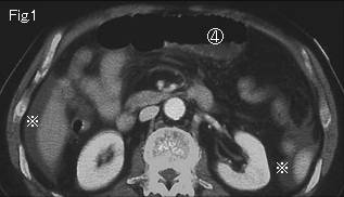
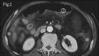
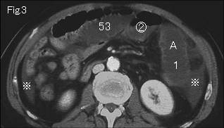
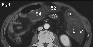
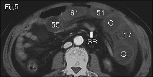
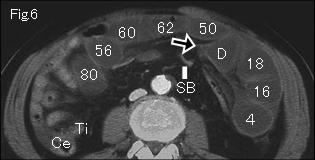
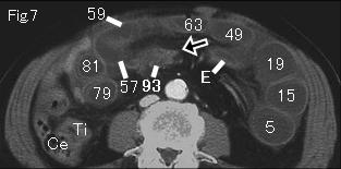
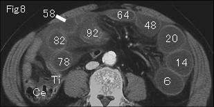
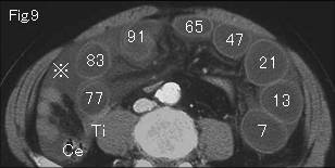
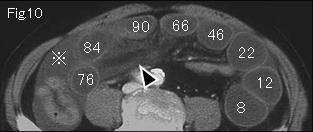
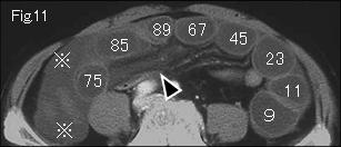
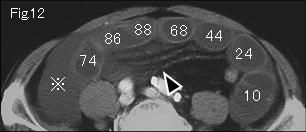
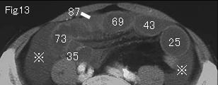
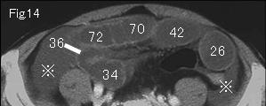
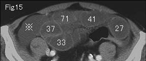
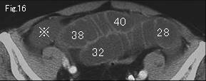
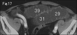
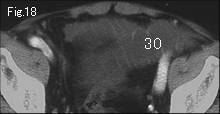
Cecum (Ce) contains no fluid and terminal ileum (Ti) in Fig.6-Fig.9 is collapsed, indicating the distended small bowel is mechanical obstruction. Gasless distension, ascites (reference mark) in Fig.9-Fig.16 and Fig.1-Fig.4, and mesenteric stranding (black arrowhead) in Fig.10-Fig.12 are highly suggestive of strangulated obstruction. A of Fig.3 occludes at E of Fig.7 manifesting a beak sign (black arrow) at D of Fig.6. 1 of Fig.3 progresses in number order, and reaches another obstruction site 93 of Fig.7 demonstrating a beak sign (black arrow). The collapsed small bowel (SB) is shown in Fig.5 and Fig.6. Circle number 4 of Fig.1 from circle number 1 of Fig.4 represent simple obstruction of cephalad side. As a result, a diagnosis of the strangulated small bowel obstruction which formes closed loop was confirmed. The wall of closed loop shows excellent contrast-enhancement, indicating no necrosis. Surgery was undertaken two days later, and transomental hernia of about 140cm small bowel with no necrosis was confirmed.
|