|
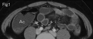
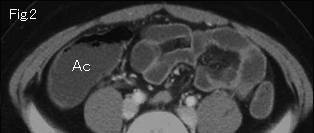
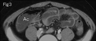
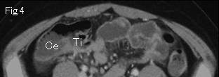
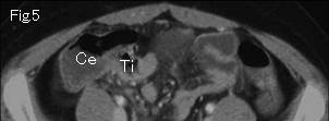
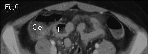
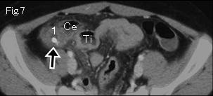
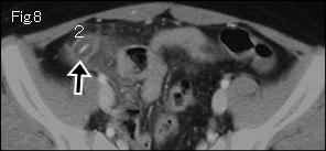
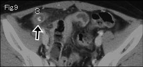
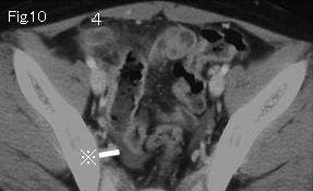
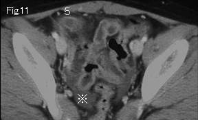
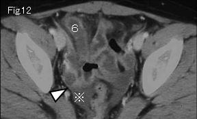
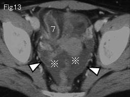
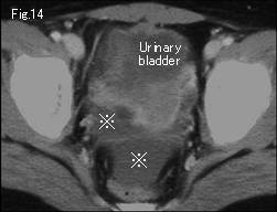
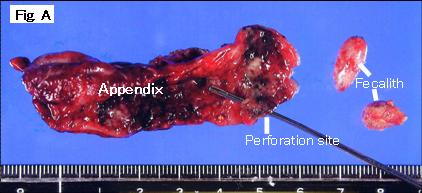
The distended small bowel in Fig.1-Fig.3, and ascending colon (Ac) and cecum (Ce) containing liquid contents in Fig.1-Fig.6 are highly indicative of paralytic ileus. Ti:Terminal ileum. If paralytic ileus is indicated, a cause of peritonitis or inflammatory disorder should be searched for. Fecalith (black arrow) in Fig.7-Fig.9 and from 1 of Fig.7 to 7 of Fig.13 indicate acute appendicitis. Paralytic ileus, ascites (reference mark) of substantial amount in Fig.10-Fig.14, and peritoneum (white arrowhead) in Fig.12 and Fig.13 showing contrast-enhancement suggestive of peritonitis, lead to diagnosis of perforated appendicitis. These findings were proved at surgery (Fig. A).
|