|
AJR Am J Roentgenol. 2007 Aug;189(2):W78-83.
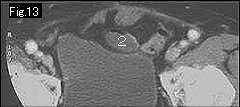
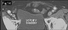
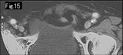
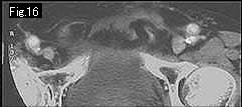
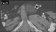
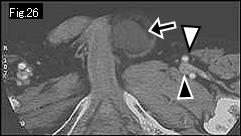
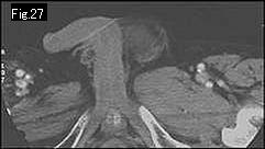
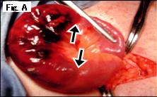
Differentiation of femoral versus inguinal hernia: CT findings.
Suzuki S, Furui S, Okinaga K, Sakamoto T, Murata J, Furukawa A, Ohnaka Y.
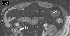
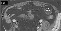
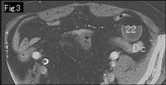
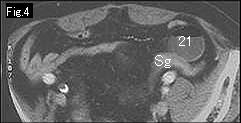
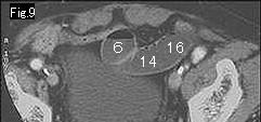
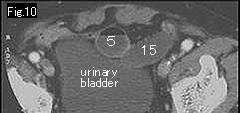
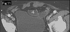
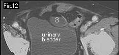
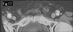
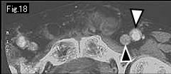
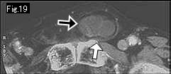
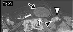
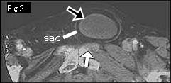
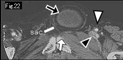
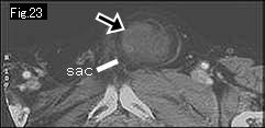
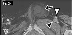
OBJECTIVE: The purpose of our study was to investigate the CT findings of femoral hernias, focusing on their differentiation from inguinal hernias. MATERIALS AND METHODS: We reviewed the records of 46 femoral hernias in seven centers (review of femoral hernias) and those of 215 groin hernias (femoral hernias, 11; inguinal hernias, 204) in one center (review of groin hernias). We evaluated the presence of hernia, extent of hernia sac based on the relationship between the hernia sac and the pubic tubercle (localized sac: sac was localized lateral to the pubic tubercle; or extended sac: sac extended medial to the pubic tubercle), and compression of the femoral vein on CT images. The chi-square test was used to assess the relationship between the CT findings and femoral versus inguinal hernias in the review of groin hernias. RESULTS: In the review of 46 femoral hernias, the lesions were detected on CT in 45. In the 45 lesions, all hernia sacs were localized, and 42 lesions showed venous compression. In the review of 215 groin hernias, all 11 femoral hernias had localized sacs with venous compression on CT. Of the 204 inguinal hernias, 98 lesions were detected on CT, 65 had extended sacs, and only 10 showed venous compression. Localized sacs with venous compression were seen much more often in the femoral hernias (11/11, 100%) than in the inguinal hernias (1/92, 1.1%) (p
|