|
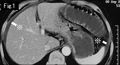
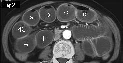
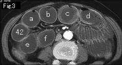
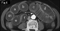
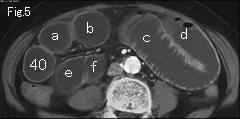
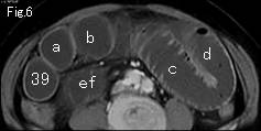
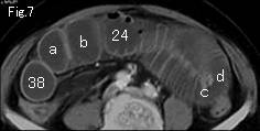
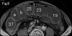
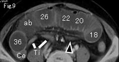
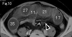
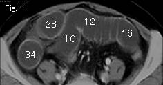
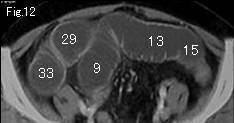
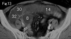
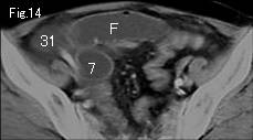
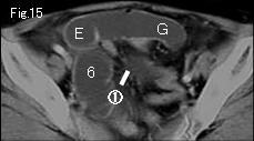
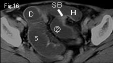
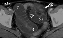
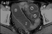
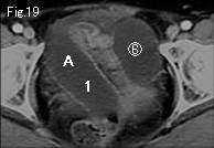
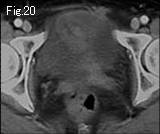
Collapsed cecum (Ce) and terminal ileum (Ti) of Fig.9, ascites (reference mark) in Fig.1, distended gasless small bowel and mesenteric stranding (black arrowhead) in Fig.9 and Fig.10, these four findings are highly indicative of strangulated obstruction. When any difficulties encountered on tracking down, the best way to solve complicated cases with many distended loops is to track down well-defined and easily-traceable loops first and put marks on. ab of Fig.9 are divided into a and b of Fig.8 and ascend as shown, c/d of Fig.7 and e/f of Fig.6 do likewise. Consequently A of Fig.19 occludes at H of Fig.16 and 1 advances to 43 of Fig.2. Now another loops of Fig.15-Fig.19 are recognized, and circle number 8 of Fig.17 from circle number 1 of Fig.15 indicate closed loop of about 15cm length. Collapsed small bowel (SB) is seen in Fig.16, and it became clear that H of Fig.16 is starting point of simple obstruction. A diagnosis of strangulated small bowel obstruction can be made, and well contrast-enhanced wall suggests viable loops. Although there was no muscle guarding, laparotomy was undertaken under accurate analysis of CT findings. At 80cm from cecum, ileum of 20cm length was incarcerated by an adhesive band forming closed loop, and revealed mild ischemic change which relieved by lysing a band.
|