|
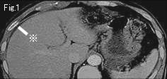
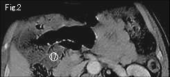
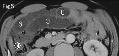
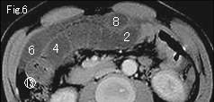
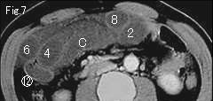
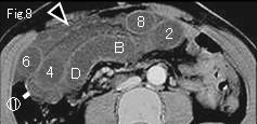
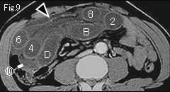
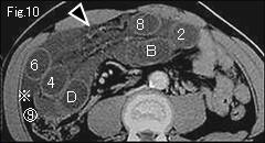
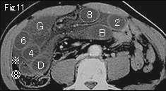
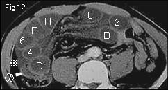
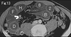
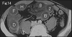
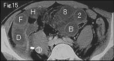
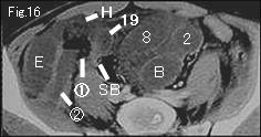
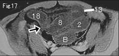
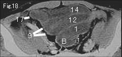
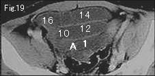
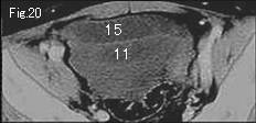
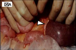
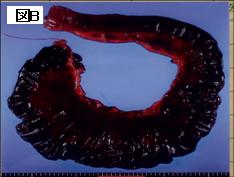
In the presence of three findings of ascites (reference mark) in Fig.1 and Fig.10-Fig.13 , gasless distended small bowel and mesenteric stranding (black arrowhead) in Fig.8-Fig.10, strangulated obstruction should be considered. Closed loop formation can be confirmed. On tracking down from A and 1 of Fig.19, A obstructs at H and 1 at 19 of Fig.16 showing beak sign (black arrow) in Fig.17. Fig.16 shows collapsed small bowel (SB), and circle number 1 of starting point of simple obstruction progresses to circle number 17 of Fig.2. Satisfactory contrast-enhancement of wall of closed loop indicates viable bowel. Ten hours later, patient was taken to surgery because of aggravated abdominal pain and peritoneal sign. As shown in Fig.A , strangulated small bowel obstruction by an adhesive band (white arrowhead) was revealed, and necrotic ileum of 90cm length (Fig.B) was excised. It is very possible that strangulated small bowel was viable at the time CT scan was taken ten hours ago.
|