|
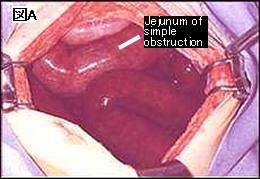
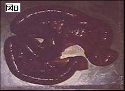
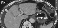
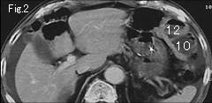
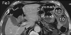
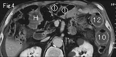
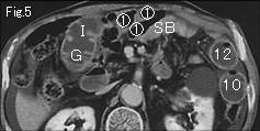
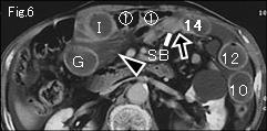
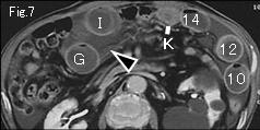
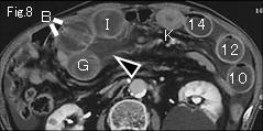
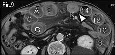
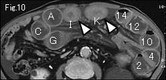
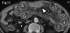
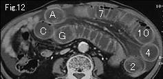
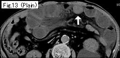
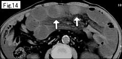
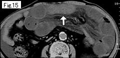
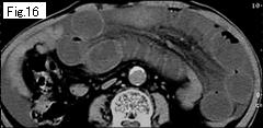
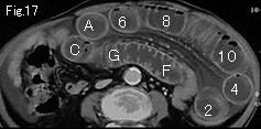
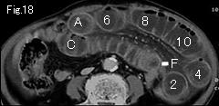
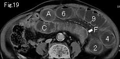
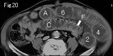
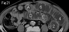
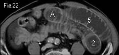
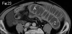
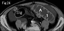
No fluid in right colon, ascites in Fig.1 (reference mark), ascites between mesentery in Fig.6-Fig.8 (black arrowhead) and gasless distended small bowel indicate that probability of strangulated small bowel obstruction is extremely high. On tracing starting with Fig.24, A and 1 occlude together at black arrow of Fig.6 indicating closed loop of length more than 100cm. Circle number 1 of Fig.6-Fig.4 are small bowels of simple obstruction, and Fig.5 and Fig.6 show collapsed small bowel (SB). Seemingly sufficient contrast-enhancement of small intestinal wall indicates no necrosis. However,I-K (white arrowheads) of Fig.9-Fig.11 show wall thickening, and strong attenuation (white arrow) on plain CT (Fig.13-Fig.16) are highly suggestive of hemorrhagic necrosis. Abdomen revealed peritoneal sign 12 hours later and surgery was performed. At 20cm from Treitz's ligament, necrotic jejunum of about 135cm length (Fig. A and Fig. B) was strangulated by adhesive band forming closed loop.
|