|
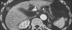
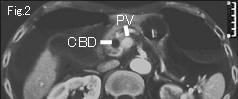
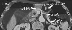
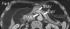
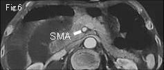
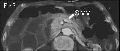
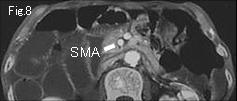
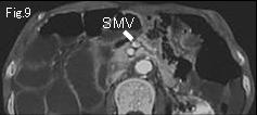
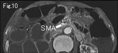
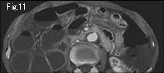
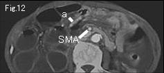
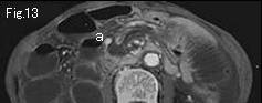
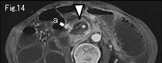
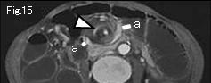
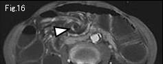
Fig.17-Fig.20 were abbreviated. CBD: common bile duct, PV: portal vein, SpA: splenic artery, SpV: splenic vein, SMA: superior mesenteric artery, SMV: superior mesenteric vein. Walls of distended small bowel group are well contrast-enhanced suggesting no necrosis or severe ischemia. However, a large quantity of ascites (reference mark of Fig.1) suggests serious pathological condition, so careful search should be made. On tracking down SMA and SMV from Fig.4, SMV does deviate to left side of SMA in Fig.4-Fig.9 and becomes unclear in Fig.10. White arrowhead of Fig.14-Fig.16 is vascular whirl sign, and it is finding to strongly suggest torsion of small intestine. a of Fig.12-Fig.15 possibly indicate twisted collapsed small intestine around SMA. Based on these findings, diagnosis of small bowel volvulus was made and immediate surgery was undertaken. Small bowel was twisted 360 degrees in clockwise direction, but there was no finding of necrosis. Because it is torsion around proximal SMA, delayed diagnosis and surgery may have caused massive small bowel necrosis and subsequent short bowel syndrome.
|