|
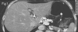
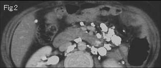
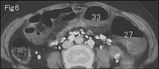
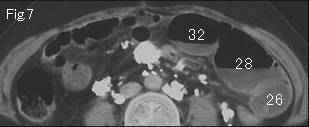
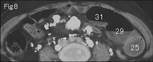
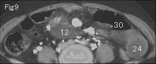
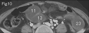
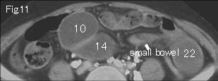
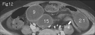
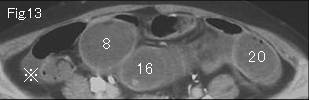
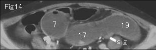
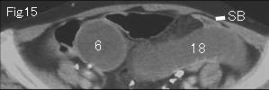
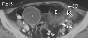
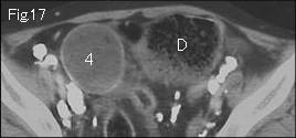
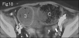
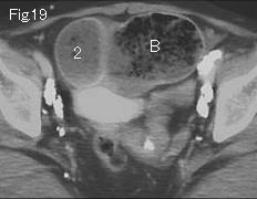
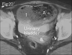
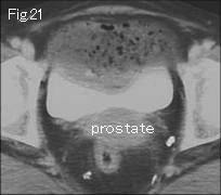
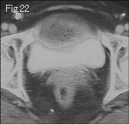
Fig.3-Fig.5 are abbreviated. Innumerable calcifications depicted in all figures are calcified mesentery and retroperitoneal lymph nodes caused by the tuberculosis peritonitis in childhood. There is no ascites around liver and in pelvic cavity, but a small quantity is present in the right lateral gutter in Fig.13 (reference mark). The wall of distended small bowel shows satisfactory contrast- enhancement and there is no mesenteric stranding to suggest strangulated obstruction. Though findings of ascites and gasless distended small bowel are in favor of strangulated obstruction, on tracking down from Fig.20, A occludes at E in Fig.16, and 1 makes progress to 33 of Fig.6 indicating simple obstruction at Fig.16. The collapsed small intestine (SB) adjacent to obstruction site is shown in Fig.15 and Fig.16, and black arrow of Fig.16 is transition zone. Because no neoplastic lesion, intussusception or compression from outside lesion are depicted, presumable cause of obstruction is by adhesion. The findings of E of Fig.16 from A of Fig.20 are referred to as ¡Èsmall bowel feces¡É (food residue), which indicates site of simple obstruction in many cases. Abdominal pain disappeared with inserted nasogastric tube for 3 days. sig:sigmoid colon.
|