|
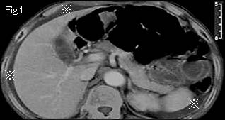
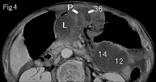
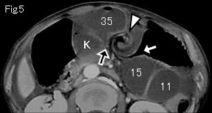
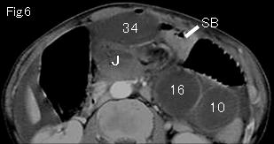
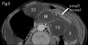
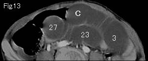
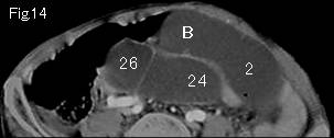
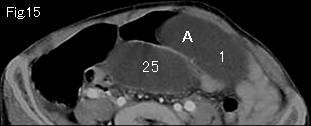
Fig. 1 shows ascites (reference marks) around liver and spleen. The distended small intestine is gasless and mural contrast-enhancement is slightly weak in comparison with viable small intestine (SB) of Fig.6 and Fig.8, and ascites between mesentery (Fig.10 and Fig. 11:black arrowheads) strongly suggest strangulated obstruction. On tracking down the distended small intestine from A and 1 of Fig.15, black arrow of Fig.5 shows beak sign, and P and 36 of Fig.4 indicate obstruction site proving closed loop formation. There is collapsed small intestine (SB) in Fig.6. A white arrow of Fig.5 presents beak sign, too, indicating starting point of oral simple obstruction. White arrowhead of Fig.5 swirls like a hurricane of weather chart in counterclockwise direction, and it is important finding called "whirl sign" indicative of volvulus (torsion). Therefore, a diagnosis of necrosis or advanced ischemia by small bowel torsion was made. Operative findings: necrotic jejunum of about 80cm with 360 degrees counterclockwise torsion.
|