|
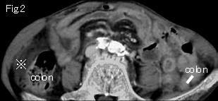
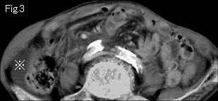
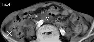
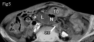
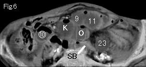
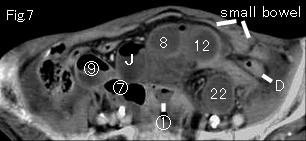
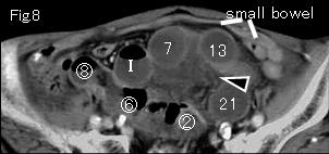
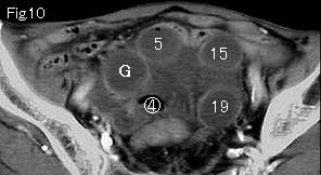
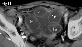
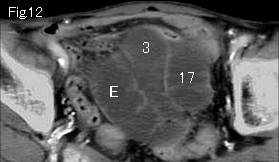
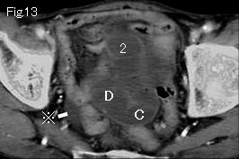
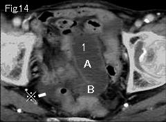
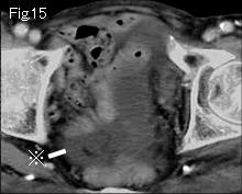
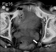
Small bowels are distended. Fig.2 shows no liquid content in right colon and collapsed left colon indicating small bowel obstruction. There is ascitic fluid (reference mark) in pelvic cavity(Fig.13-Fig.16) and right lateral gutter(Fig.2-Fig.4). Fig.8 and Fig.9 show mesenteric stranding (black arrowheads), and distended small intestines are gasless, suggesting high possibility of strangulated obstruction. Tracking down 1 and A of Fig.14 to caudal side arrives at obstruction site of 23 and O of Fig. 6 which indicates closed loop formation. Collapsed small intestine (SB) is seen in same Fig.6 and simple obstruction starts from circle number 1 of FIg.7. Attenuation of wall of closed loop slightly decreases in comparison with collapsed small bowel of Fig.7 and Fig.8 suggesting ischemia. D:descending colon. CT diagnosis of strangulated small bowel obstruction was made, and operation was performed. Necrotic small intestine of about 45cm length formed closed loop by an adhesive band associated with mild torsion.
|