|
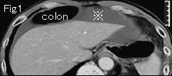
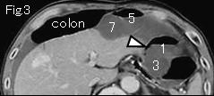
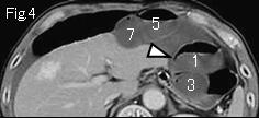
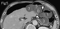
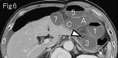
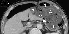
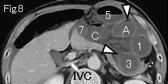
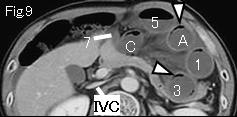
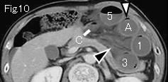
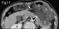
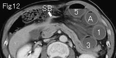
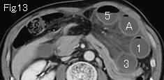
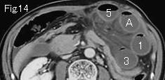
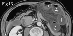
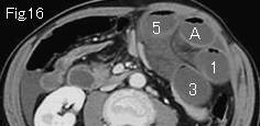
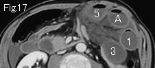
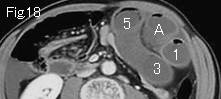
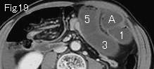
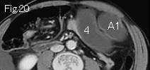
The wall of dilated and gasless small bowel shows poor contrast enhancement. There is ascites in Fig.1(referencr mark) and black arrowheads of Fig.10 and Fig.11 depict obvious stranding of mesentery suggesting strangulated bowel obstruction. A of Fig.20 occludes at C of Fig.10, and 1 of Fig.20 occludes at 7 of Fig.9. There is the collapsed small bowel (SB) in Fig.11 and Fig.12 indicating that 1-7 of Fig.9 and A-C of Fig.10 form closed loop. Intramural gas of closed loop (the linear gas along wall which does not form air-fluid level: white arrowhead) in Fig.3-Fig.10 indicates necrosis or ischemia. A diagnosis of small bowel necrosis of estimated 100cm length by strangulated bowel obstruction can be made. Large bowel gets from liver front into subphlenic space, and this is called ¡ÈChilaiditi's syndrome¡É. IVC of Fig.8 and Fig.9 is collapsed suggesting strong dehydration. Surgery showed strangulated bowel obstruction by an adhesive band and 80cm necrotic ileum.
|