|
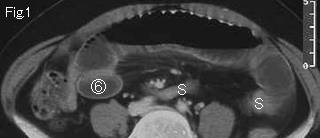
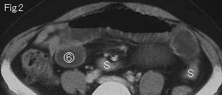
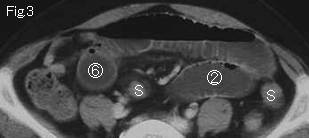
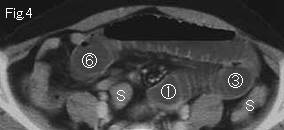
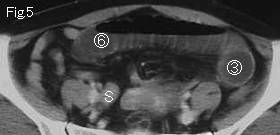
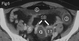
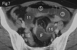
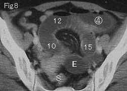
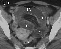
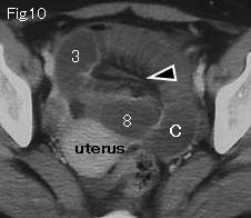
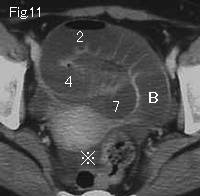
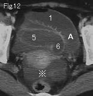
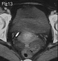
The distension limited to small bowel indicates small bowel obstruction. There is ascites (reference mark) in Fig.11-Fig.13, the dilated small bowel in pelvic cavity is gasless, Fig.10 shows mesenteric fluid or fat stranding (black arrowhead). These three findings suggests strangulated bowel obstruction. Therefore attention should be paid to the bowel group with mesenteric stranding in pelvic cavity, and tracing from A and 1 of Fig.12 should be attempted to prove closed loop formation. Both A and 1 obstruct at G and 17 of Fig.6 , there is nearby collapsed small bowel (SB). Distended bowels from circle number 1 of Fig.4 to circle number 6 of Fig.1 are loops of simple obstruction. Accordingly, closed loop formation of about 50cm length was confirmed. No thickening of wall of closed loop and satisfactory contrast enhancement indicate no necrosis. S:sigmoid colon. CT findings were diagnosed accurately and patient was taken to surgery. There was an adhesive band between left ovary and peritoneum with which about 45cm ileum was strangulated, but found to be viable. Severance of band was all necessary.
|