|
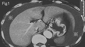
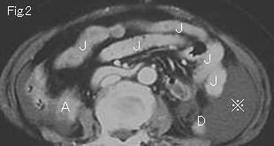
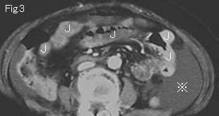
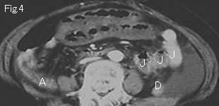
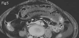
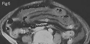
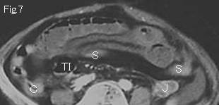
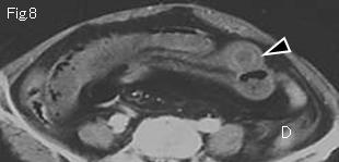
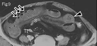
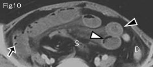
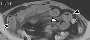
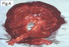
There is a large quantity of ascites in Fig.1-Fig.3 (reference mark). Wall of jejunum (J), right colon (A: ascending colon, C: cecum), TI (terminal ileum), D (descending colon) and S (sigmoid colon) of Fig.2-Fig.11 is well contrast-enhanced. In contrast with those, wall of distended small bowel shows poor or no enhancement indicating necrotic or highly ischemic bowel. Bubbly gas (black arrow) of Fig.9-Fig.11 could be intraluminal gas situated between Kerckring folds, but white arrowhead of Fig.10 and Fig.11 is curvilinear without forming air-fluid level is intramural pneumatosis. Some small bowels present wall thickening ( >3mm, Fig.8-Fig.11: black arrowhead). Laparotomy revealed necrotic ileum of about 120cm length with strangulation caused by adhesive band (Fig.A).
|