|
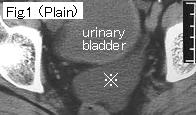
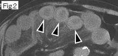
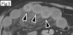
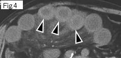
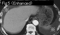
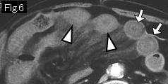
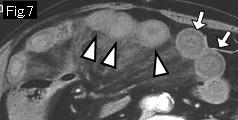
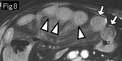
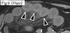
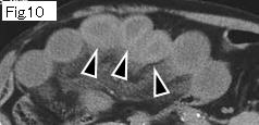
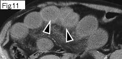
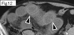
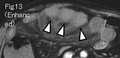
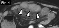
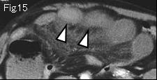
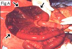
There is massive ascites in pelvic cavity of Fig.1 (reference mark), around liver and spleen of Fig.5. Small bowel of white arrowhead(Fig.6-Fig.8 and Fig.13-Fig.15) appears to show strong enhancement on contrast-enhanced CT indicating viable wall. However, noncontrasted CT (black arrowhead of Fig.2-Fig.4 and Fig.9-Fig.12) demonstrates high density (equivalent to or more than muscle density), indicating wall thickening by hematoma, which strongly suggests hemorrhagic necrosis. Small bowel of white arrow of Fig.6-Fig.8 shows wall thickening by submucosal edema. Surgery revealed strangulated obstruction by adhesive band, and hemorrhagic necrosis of jejunum (Fig.A, black arrow).
|