|
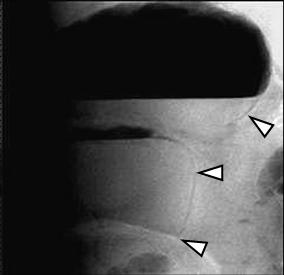
CT findings of bowel necrosis are, 1) poor or absent enhancement of bowel wall on intravenously contrast-enhanced CT, 2) increased attenuation of bowel wall on non-contrast CT (hemorrhagic necrosis), 3) free air, 4) gas in portal vein or SMV, 5) pneumatosis (intramural gas), 6) wall thickening with or without target sign, 7) ascites, 8) thickening of peritoneum, mesentery or retroperitoneal fascia adjacent to bowel wall. Specificity is high in 1, 2 and 5.
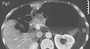
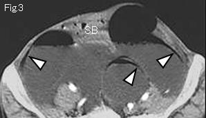
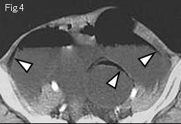
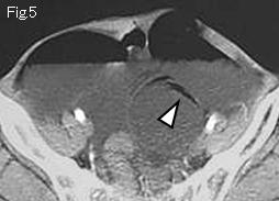
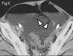
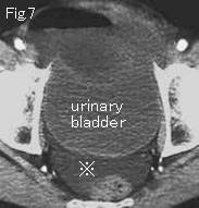
There is ascites in Fig.1 and Fig.7 (reference mark). Dilated (>2.5cm) small intestine shows lack of contrast enhancement of the wall, or only slightly enhanced compared with rectum (R), sigmoid colon (S) of Fig. 6 and other small intestines (SB) of Fig.2 and Fig.3. Furthermore, there is the linear gas (white arrowheads) along the wall without forming niveau (air-fluid level) indicating pneumatosis (intramural gas) due to small bowel necrosis. Surgery revealed small bowel necrosis caused by strangulated small bowel obstruction with adhesion and torsion.
|