CT of Acute Abdomen

Epigastric Pain series 3 RESIDENT COURSE Answer [ER Case 12]

Perforated gastric ulcer


There is free air (white arrowheads) in Fig.2-Fig.4. Fig.8 and Fig.9 show ascites (reference mark) in the Morison pouch. The lesser curvature of stomach demonstrates edematous wall thickening (black arrowheads) from Fig.2 and extends to anterior wall of gastric body of Fig.4-Fig.6, and to posterior wall of antrum of Fig.7 and Fig.8. Black arrow of Fig.3-Fig.6 is a wall defect and a diagnosis as perforation of an acute ulcerative lesion of anterior wall of gastric body can be made. The finding was confirmed at surgery (Fig.A).


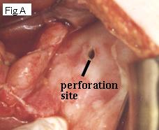
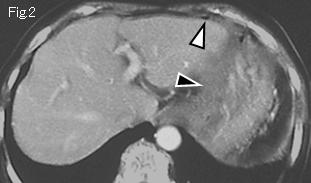
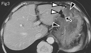
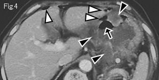
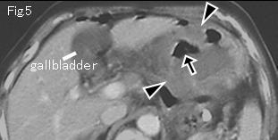
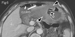
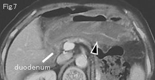
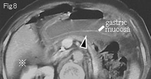
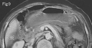
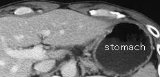
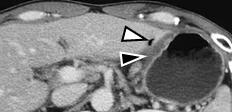
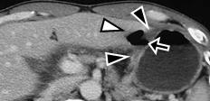
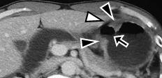
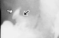
Reference case (perforated gastric ulcer): A 38-year-old male presented with sudden onset of severe epigastric pain for several hours. Temperature: 38.6 degrees Celsius. Physical examination revealed localized tenderness and board-like rigidity in epigastrium. Stomach shows edematous wall thickening (black arrowhead) , white arrowhead is free air, and black arrow is an acute ulcerative lesion. Gastrografin contrast study showed an ulcer (black arrow) with localized leakage (white arrow) of contrast medium without spreading into free peritoneal cavity. Patient made full recovery with conservative treatment. Above finding was confirmed by endoscopy and biopsy reported as benign ulcer.


 Previous Case    Next Case   to Questions Page   to Practical Exercises Page