CT of Acute Abdomen

Epigastric Pain series 2 RESIDENT COURSE Answer [ER Case 7]

Perforated gastric ulcer


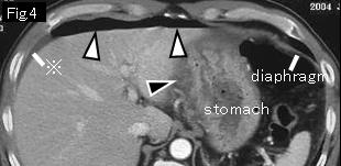
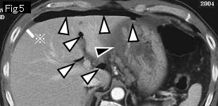
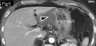
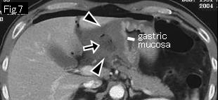
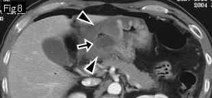
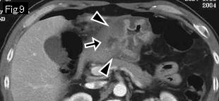
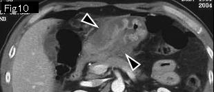
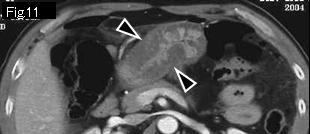
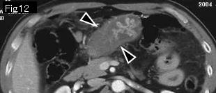
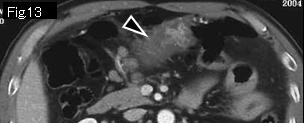
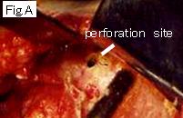
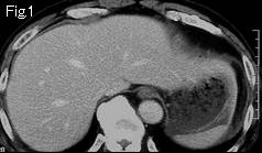
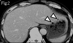
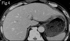
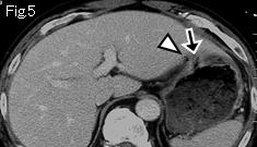
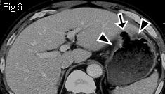
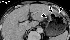
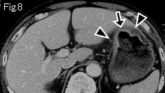
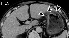
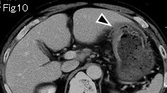
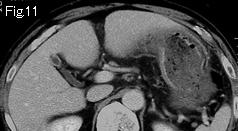
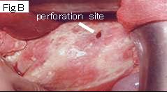
There are free air (white arrowheads) and ascites (reference mark) in Fig.4 and Fig.5, and likelihood of gastrointestinal perforation is extremely high. The wall of gastric angle begins to thicken by submucosal edema from Fig.4 (black arrowhead) and extends to entire circumference of antrum from Fig.10. Because black arrow of Fig.7-Fig.9 indicates an acute ulcerative lesion spreading through gastric angle to anterior wall of antrum, a diagnosis of gastric ulcer perforation can be made. In Fig.7, base of deep ulcer (black arrow) of gastric angle is recognized at remote site from gastric mucosa. Surgery revealed a perforation of 3 by 2cm size at lesser curvature of the antrum (Fig.A), and partial gastrectomy was undertaken.


Reference Case (perforated gastric ulcer): A 63-year-old male who had gastric ulcer 4 years ago, presented with aggravating epigastric pain which started 3 hours ago after dinner. Temperature: 35.5 degrees Celsius. Examination showed tenderness and rebound tenderness in epigastrium.
White arrowheads of Fig.2 and Fig.5 is very likely to be free air. There is edematous wall thickening of anterior wall of body of stomach in Fig.6-Fig.10 (black arrowheads) and black arrow of Fig.5-Fig.9 is an acute ulcerative lesion, which indicates gastric ulcer perforation which was confirmed at surgery (Fig.B).


 Previous Case    Next Case   to Questions Page   to Practical Exercises Page