|
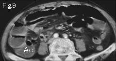
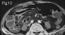
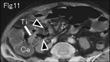
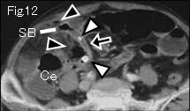
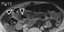
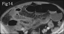
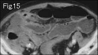
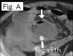
Fig.1-Fig.8 are abbreviated. Small bowel is distended, however, ascending colon (Ac of Fig.9 and Fig.10) and cecum (Ce of Fig.11-Fig.13) include liquid contents similar to distended small bowel, which indicates high possibility of paralytic ileus instead of mechanical obstruction. Black arrowheads of Fig.11-Fig.13 are extraluminal free air, black arrows of Fig.12 is indicative of collapsed appendix including two fecalith (white arrowheads). Based on these findings, diagnosis of perforated appendicitis can be made. C: cecum, Ti: terminal ileum. After one week's conservative treatment, an abscess was formed in right lower quadrant (white arrow of Fig. A). Above findings were confirmed at surgery.
|