|
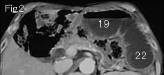
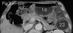
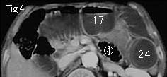
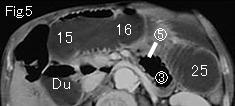
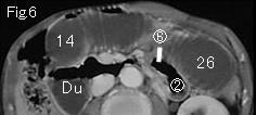
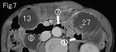
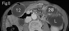
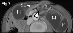
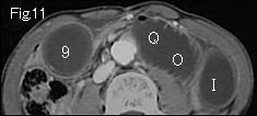
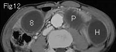
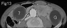
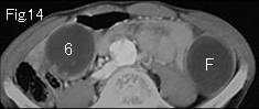
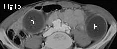
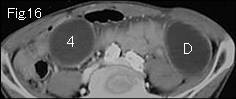
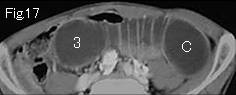
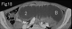
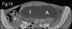
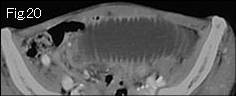
Distension of limited to small bowel and no fluid content in right colon (Fig.17-Fig.20) indicates mechanical obstruction of small bowel. There is no ascites, and wall of distended small bowel is well contrast-enhanced. Distended small bowels are gasless suggesting high possibility of strangulated obstruction. White arrowhead of Fig.9 shows "whirl sign" of mesentery and intestine indicating torsion. Because confirmation of closed loop formation leads to a definite diagnosis of small bowel volvulus, distended bowel should be tracked down. 1 of Fig.19 occludes at 28 of Fig.8 and A at P of Fig.10 indicating closed loop formation. Fig.9 and Fig.10 demonstrate collapsed small bowel (SB), and simple obstruction starts at circle number 1 of Fig.7 connecting to duodenum (Du of Fig.6). Accurate CT diagnosis of jejunal volvulus was made and immediate surgery revealed 100cm jejunum twisted 360 degrees in clockwise fasion at 20cm from Treitz's ligament. Patchy subserosal ecchymosis and mildly ischemic findings were found, but improved immediately by detorsion.
|