|
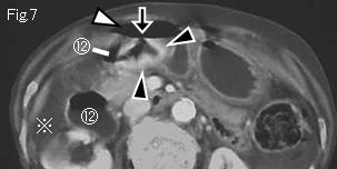
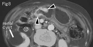
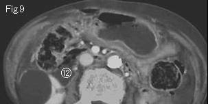
There are free air (white arrowheads) and ascites (reference mark). Comparing with the other part, the wall of antrum in Fig.7 and Fig.8 is strongly contrast-enhanced (black arrowheads) and highly suggestive of a neoplasmic lesion. Free air (white arrowhead) adjacent to perforation site (black arrow) in Fig.7 is also depicted, and it is very likely that a cause of gastrointestinal perforation is a gastric cancer. Circled number 12: duodenum. Surgery revealed perforated firm tumor in anterior wall of antrum that was consistent with gastric cancer.
|
|
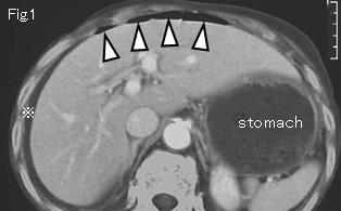
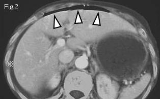
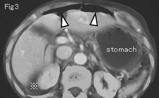
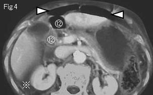
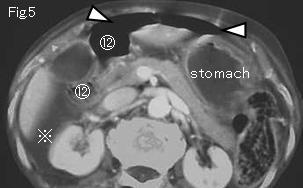
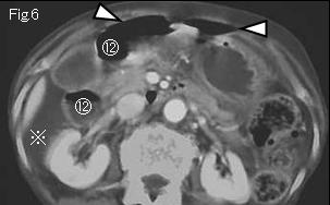
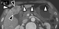
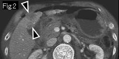
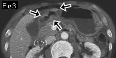
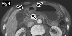
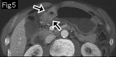
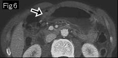
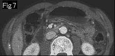
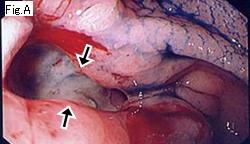
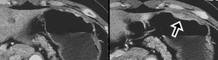
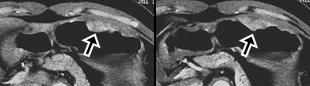
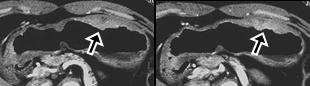
Reference case 1 (gastric cancer perforation): A 63-year-old male. Gradually increasing epigastric pain developed three hours ago. Temperature: 37.4 degrees Celsius, abdominal examination showed signs of peritonitis in the entire abdomen. CT images show free air (white arrowhead) in Fig.1, and black arrowheads of Fig.1 and Fig.2 suggests a metastatic lesion. Black arrows of Fig.3-Fig.6 show wall thickening slightly strongly, irregularly contrast-enhanced in comparison with gastric wall of the other part, suggesting gastric cancer. The perforated gastric lesion was confirmed by surgery, and gastric cancer was diagnosed by endoscopy (Fig.A: black arrow).
|
|
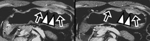
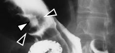
Reference case 2 (5mm slice, non-perforated gastric cancer): A 47-year-old female. On mass examination, abnormal findings were pointed out by upper GI series. Black arrows of CT images is a well-enhanced cancerous lesion with ulcer (white arrowhead), and upper GI series (prone position) shows a cancer (black arrowhead) associated with ulcer (white arrowhead) in anterior wall of body of stomach. Biopsy revealed adenocarcinoma.
|
|