CT of Acute Abdomen

Epigastric Pain series 3 RESIDENT COURSE Answer [ER Case 13]

Perforated duodenal ulcer


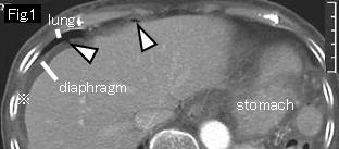
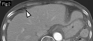
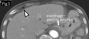
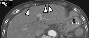
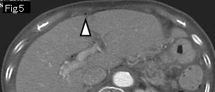
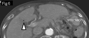
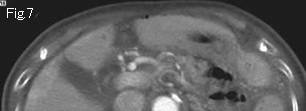
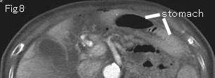
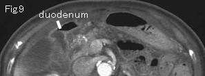
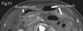
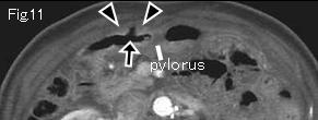
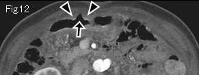
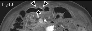
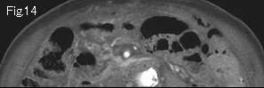
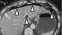
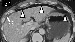
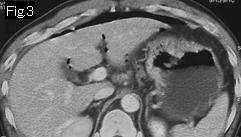
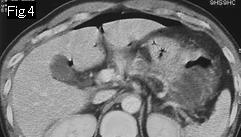
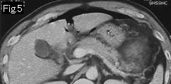
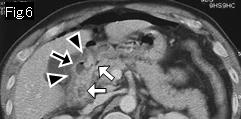
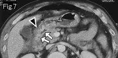
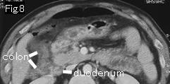
Fig.1-Fig.6 show free air (white arrowheads), and there is a little ascites (reference mark) in Fig.1-Fig.4. Thickened-appearing wall of stomach of Fig.1-Fig.6 is not abnormal findings because it is collapsed. No submucosal edema is seen in wall of stomach. Black arrowheads of Fig.11-Fig.13 present slight mural thickening of anterior wall of duodenal bulb, and black arrow of Fig.11-Fig.13 is an acute ulcer, which was confirmed at surgery. However, likelihood to succeed with conservative treatment is very high because of small amount of ascites and short period from the onset of pain.


Reference case (duodenal ulcer perforation): A 47-year-old male presented with epigastric pain that developed suddenly several hours ago. Temperature: 37.2 degrees Celsius. Abdomen showed peritoneal signs and board-like rigidity.
There are free air (white arrowheads) and ascites (reference mark) in Fig.1 and Fig.2. Duodenal bulb shows edematous wall thickening (black arrowhead) in Fig.6 and Fig.7. White arrow indicates duodenum lumen, and a defect (black arrow) in edematous wall thickening of Fig.6 is an acute ulcer. Laparotomy revealed perforated ulcer of anterior wall of duodenal bulb.


 Previous Case    Next Case   to Questions Page   to Practical Exercises Page