CT of Acute Abdomen

Epigastric Pain series 2 EXPERT COURSE Answer [EE Case 6]

ADML(acute duodenal mucosal lesion)


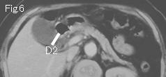
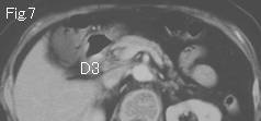
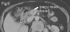
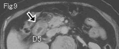
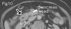
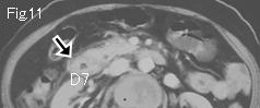
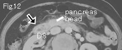
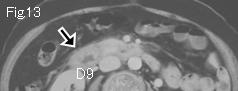
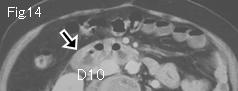
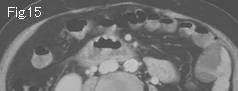
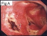
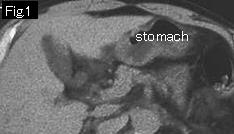
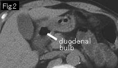
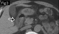
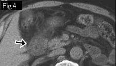
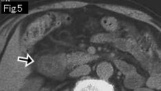
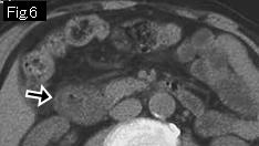
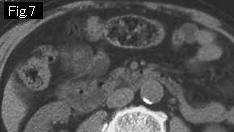
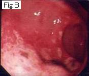
D1 in Fig.5 is duodenal bulb, and D3-D9 are descending portion. D5 of Fig.9-D10 of Fig.14 demonstrate wall thickening (black arrow) by submucosal edema. Absence of pancreatic head enlargement, neighboring inflammatory findings, or heterogeneity of pancreas parenchyma indicate possibility of pancreatitis is unlikely. It is acute inflammatory lesion localized in descending portion of duodenum, namely acute duodenitis (ADML: acute duodenal mucosal lesion). Endoscopy (Fig.A) showed multiple shallow acute ulcers associated with acute duodenitis and clot in descending portion of duodenum.


Reference Case (Plain CT, ADML): A 77-year-old male developed epigastric pain 11 hours ago, and was taken to ER because abdominal pain increased over past five hours. Temperature: 36.8 degrees Celsius. Examination showed tenderness in epigastrium.
CT images show wall thickening (black arrow) by submucosal edema from duodenal bulb of Fig.3 to descending part of Fig.6 . Endoscopy on the next day revealed redness, edema and erosions in descending duodenum indicating ADML (Fig.B).


 Next Case   to Questions Page   to Practical Exercises Page