|
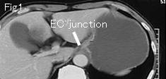
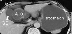
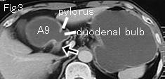
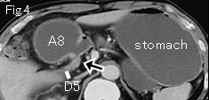
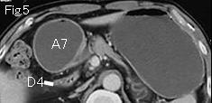
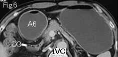
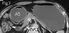
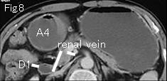
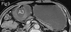
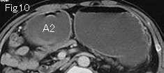
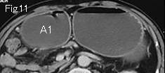
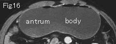
Fig.12-Fig.15 are abbreviated. Stomach is distended. A1 of Fig.11 (antrum of stomach) advances in number order to caudal side, ending at A10 of Fig.2, which indicate black arrow of Fig.3 and Fig.4 is the site of occlusion. On tracking back from duodenum D1 of Fig.8 to caudal side, D1 arrives at the occlusion site of D5 of Fig.4. There is no surrounding lesion to compress, and the site of occlusion (black arrow) does not show the typical wall thickening due to neoplasm, which leads to a diagnosis of obstructive duodenal ulcer. Collapsed IVC of Fig.6 and Fig.7 indicates severe hypovolemia. Endoscopy two days later showed an active ulcer and stricture by duodenal ulcer scar.
|