|
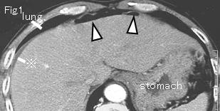
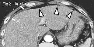
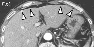
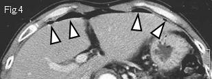
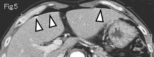
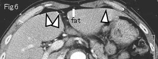
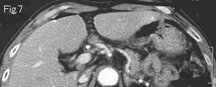
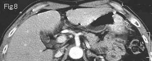
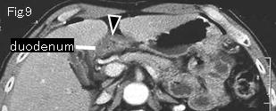
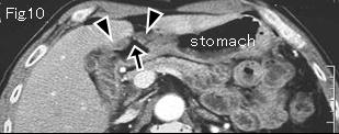
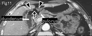
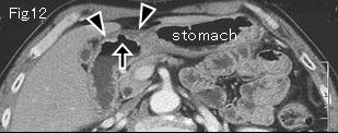
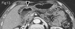
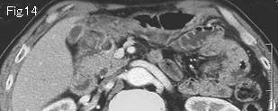
Fig.1 and Fig.2 show a little ascitic fluid (reference mark), and white arrowheads in Figs.1-Fig.6 represent free air. Because of absence of any gastric wall edematous thickening, the duodenum should be searched for next. Black arrowheads (Fig.9–Fig13) show edematous wall thickening of anterior wall of duodenal bulb. Black arrows (Fig.10–Fig12) indicate a wall defect highly suggestive of an acute duodenal ulcer perforation. Because of gradual improvement of abdominal pain and the presence of only small quantity of ascitic fluid on CT, conservative treatment was undertaken, which succeeded without complications. Endoscopy on fifth day revealed a sealed-off active ulcer in anterior wall of duodenal bulb.
|