CT of Acute Abdomen

Epigastric Pain series 1 EXPERT COURSE Answer [EE Case 5]

Perforated duodenal ulcer


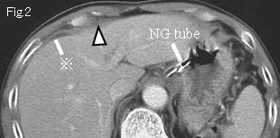
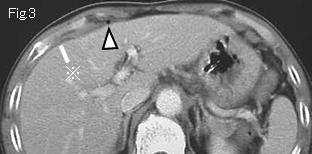
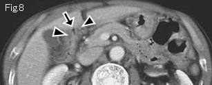
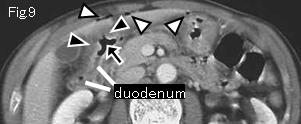
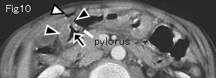
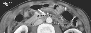
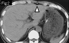
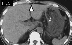
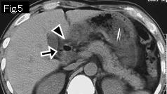
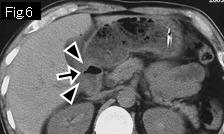
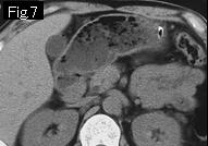
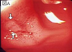
Both Fig.2 and Fig.3 show free air (white arrowhead) and ascitic fluid (reference mark). The anterior wall of duodenal bulb (black arrowheads of Fig.8-Fig.10) is thickened by submucosal edema, and black arrows indicate a duodenal ulcer. Fig.9 and Fig.10 show free air (white arrowheads) anterior to duodenal ulcer and beneath anterior abdominal wall, which are also highly suggestive of perforated duodenal ulcer. Laparotomy revealed a perforated ulcer (8mm in size) in anterior wall of duodenum.


Reference Case (Plain CT, perforated duodenal ulcer): A 47-year-old male came to the emergency room complaining of epigastric pain that had started several hours before. Body temperature: 37.0 degrees Celsius. Abdomen was soft and flat with tenderness in epigastrium.
There is free air (white arrowheads) but no ascitic fluid. Because of absence of any findings to suggest acute lesions in stomach, attention should be paid to the duodenum. Fig.5 and Fig.6 show edematous wall thickening (black arrowheads) of the duodenal bulb and a duodenal ulcer is depicted by a small little gas (black arrow). Endoscopy (Fig.A) showed an active ulcer (white arrows) that was already sealed off.


 Previous Case   to Questions Page   to Practical Exercises Page