|
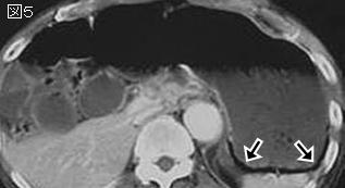
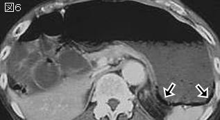
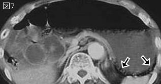
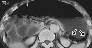
文献考察1):壁内気腫(intramural gas,pneumatosis cystoides intestinalis)
AJR Am J Roentgenol. 2003 Mar; 180(3):733-6.
Pneumatosis intestinalis in patients with ischemia: correlation of CT findings with viability of the bowel.
Kernagis LY, Levine MS, Jacobs JE.
OBJECTIVE: The purpose of our study was to reassess the CT finding of pneumatosis in intestinal ischemia to determine whether it indicates transmural necrosis versus partial mural ischemia and also to determine whether other CT findings can be used to predict which patients with pneumatosis are likely to have viable bowel. CONCLUSION: The CT finding of pneumatosis does not always indicate transmural infarction of the bowel in intestinal ischemia. Patients with associated portomesenteric venous gas are more likely to have transmural infarction than those with pneumatosis alone.PMID: 12591685
追記:壁内気腫には2種類あり,primary(pneumatosis cystoides intestinalis:15%)とは原因不明の,気泡状の嚢胞性の気腫で,良性で症状がなく通常随伴する病変なく偶然発見される.secondary(85%)は線状か全周性のことが多く,種々の原因で二次的に発生するもので,腸管粘膜を損傷する状態(腸管壊死,腸管虚血,腸閉塞,潰瘍,悪性腫瘍,腸炎,腸管吻合部周辺,化学療法や大量のステロイド投与時)に内圧上昇が加われば起こりうるものである.
15例中60%は腸管壊死あり,40%は壊死を伴わない壁内気腫で虚血状態だがreversibleであった.同時に門脈内ガスがあれば100%壊死,壁内気腫と他の虚血所見があり門脈内ガスを伴わなければ56%が壊死であった.
文献考察2):胃壁内気腫(gastric emphysema)
症例に学ぶ Gastric Emphysemaの1例
Author:岩泉守哉(県西部浜松医療センター), 山田正美, 北川陸生, 竹平安則, 花島一哲, 室久剛, 川村素子, 岩岡泰志, 和田朋彦, 森田悟, 川田一仁
Source:日本消化器病学会雑誌(0446-6586)99巻6号 Page642-645(2002.06)
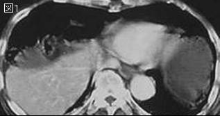
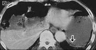
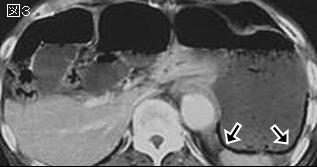
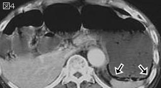
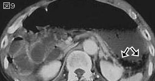
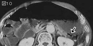
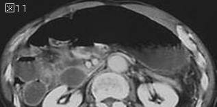
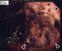
Abstract:86歳男.1日3から4回の嘔吐を認め,絶食で経過観察していたが改善しないため,救急車で搬送された.腹部単純CTで胃内に内容物及びガスの貯留と,胃底部から前庭部の後壁内を中心に線状のガス像を認めた.嘔吐が頻回であったため入院の上経鼻胃管を挿入し,補液と絶食で経過観察した.胃内容物は第2病日には多量に排出され嘔吐も消失,第5病日の腹部X線写真で胃壁内ガス像の消失を確認した.上部消化管内視鏡検査では十二指腸球部に潰瘍瘢痕を多数認め,変形が著明で内視鏡の通過に難渋した.更に潰瘍瘢痕口側にヘマチンの付着した活動期の潰瘍を認めた.胃粘膜には明らかな異常は認められなかった.obstructive typeのgastric emphysemaと診断した.十二指腸潰瘍はH2受容体拮抗薬とアルギン酸ナトリウムの内服で観察し,上部消化管内視鏡検査では潰瘍の治癒傾向を確認した.
追記:胃の壁内にガスが認められる病態は,A:gastric emphysema,B:emphysematous gastritis,C:cystic pneumatosisの3つに分類される.AはX線写真で胃壁内に沿った線状ガス像を呈するもので,ガス産生菌を起因としないものである.Bはガス産生菌により発症し,臨床経過として発熱,強い上腹部痛,悪寒,白血球増加が認められ,X線写真で泡状のガス像が認められる.Cはpneumatosis cystoides intestinalisの胃病変で,X線写真で胃壁内に嚢胞状(1-2mmのブドウの房状)にガスが認められるものである.gastric emphysemaはさらに,1)traumatic type,2)obstructive type,3)pulmonary typeに分類される.traumatic typeは胃切除,胃瘻造設術などで粘膜の欠損,断裂より生じる.obstructive typeは幽門狭窄などで内圧が上昇し,さらに粘膜障害が存在することでその部位から胃壁内へガスが侵入するものである.pulmonary typeは基礎疾患として慢性閉塞性肺疾患があり,ブラの破裂で気管支周囲から縦隔に漏出したガスが腹膜腔に沿って胃の漿膜下に到達し生ずる.本邦報告例51例中obstructive typeが最も多く72%,traumatic typeが24%,pulmonary typeは4%と少なかった.obstructive typeのうち,悪性腫瘍によるものは43%,良性潰瘍によるものが19%を占めていた.
文献考察3):気腫性胃炎(emphysematous gastritis)と胃壁内気腫(gastric emphysema)
Radiographics. 1996 Sep;16(5):1035-54.
CT of the stomach: spectrum of disease.
Fishman EK, Urban BA, Hruban RH.
In evaluation of gastric disease, computed tomography (CT) has proved to be a valuable adjunct to barium studies and endoscopy. CT clearly demonstrates the primary pathologic condition and shows extension of disease to adjacent or distant structures. Useful in staging gastric cancer, CT has also proved valuable in detecting and defining the extent of other gastric neoplasms such as lymphoma, leiomyosarcoma, and metastasis to the stomach. Recent advances in CT technology such as spiral CT-coupled with air contrast gastric studies and a better understanding of the need to optimize CT protocols-suggest that the value of CT in these applications will increase. CT has also been shown to be valuable in detection and differentiation of other gastric conditions such as benign tumors, Helicobacter pylori and other infections, various forms of gastritis (radiation, eosinophilic, and emphysematous), ulcer disease, Menetrier disease, and varices. Adequate gastric distention is essential for successful gastric CT.PMID: 8888389
追記:気腫性胃炎は感染性の胃炎で,死亡率が60-80%と高く,重篤な疾患である.ガス産生性菌による胃壁の感染で,胃壁内にガスを認める.肥厚した胃壁内に散在する気泡状のガス像が特徴で,他方gastric emphysemaは壁肥厚のない胃壁に線状のガス像を特徴とするが,鑑別は困難なこともある.
文献考察4):気腫性胃炎(emphysematous gastritis)と胃壁内気腫(gastric emphysema)
Radiographics. 2003 Jan-Feb;23(1):75-87.
Current role of CT in imaging of the stomach.
Horton KM, Fishman EK.
Recent advances in computed tomographic (CT) technology and three-dimensional (3D) imaging software have sparked renewed interest in using CT to evaluate gastric disease. Multidetector row CT scanners allow thinner collimation, which improves the visualization of subtle tumors as well as the quality of the 3D data sets. When water is used as an oral contrast agent, subtle disease is easier to visualize, especially when a rapid contrast material bolus is intravenously administered. Adenocarcinoma is the most common gastric malignancy and typically appears as focal or segmental wall thickening or a discrete mass. Gastric lymphoma can have a CT appearance similar to that of adenocarcinoma. Both gastric adenocarcinoma and lymphoma may be associated with adenopathy. Gastrointestinal stromal tumors (GISTs) tend to appear as well-defined masses that arise from the gastric wall and may be exophytic when large. GISTs are usually not associated with significant adenopathy. In addition to gastric malignancies, CT can also help detect inflammatory conditions of the stomach, including gastritis and peptic ulcer disease. CT angiography is especially helpful for depicting the gastric vasculature, which may be affected by a variety of disease conditions. PMID: 12533643(full text)
追記:emphysematous gastritisは肥厚した壁内のガス像を特徴とするが,gastric emphysemaと同様なガス像を示すこともある.前者は生命を脅かす重篤な疾患であり,後者は無症状で,胃壁内ガス像は自然に消失する.
|