|
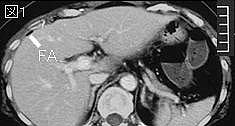
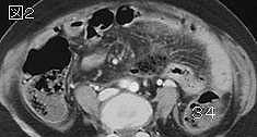
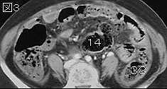
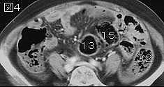
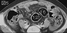
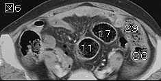
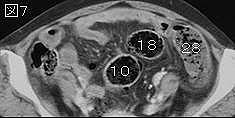
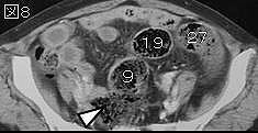
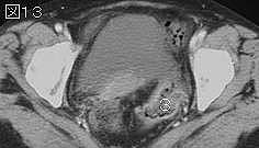
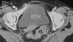
文献考察:憩室炎の診断,合併症の診断,治療方針の決定にCTは極めて有用
Ambrosetti P, Becker C, Terrier F.
Colonic diverticulitis: impact of imaging on surgical management -- a prospective study of 542 patients.
Eur Radiol. 2002 May;12(5):1145-9.
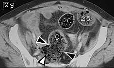
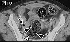
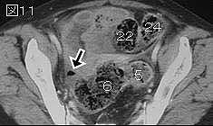
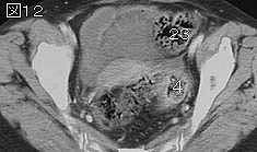
The aim of this study was to compare the performance of the CT and the water-soluble contrast enema (CE) in the diagnosis and the severity of acute left-colonic diverticulitis, and to recognize the impact of CT during the acute phase and after a first acute episode successfully treated medically. From 1986 to 1997, all patients admitted in our emergency center with clinically suspected left-colonic diverticulitis had a CE and a CT within 72 h of their admission, unless clinical findings required immediate laparotomy. They were prospectively included in the study if one or both radiological exams showed signs of acute diverticulitis and/or diverticulitis was surgically removed and histologically proven. Diverticulitis was considered moderate when CT showed localized thickening of the colonic wall (5 mm or more) and inflammation of pericolic fat and CE showed segmental lumen narrowing and tethered mucosa; it was considered severe when abscess and/or extraluminal air and/or contrast were observed on CT and when one or both of the latter signs were seen on CE. Five hundred forty-two patients entered the study; 465 patients (86%) had a CT exam, 439 (81%) had a CE, and 420 (77%) had both exams. The performance of CT is significantly superior to CE in terms of sensitivity (98 vs 92%, p
|