|
文献考察:アメーバ性肝膿瘍
【外科エンサイクロペディア】 アメーバ性肝膿瘍(解説/特集)
Author:安保義恭(北海道大学 腫瘍外科), 近藤哲, 加藤紘之
Source:外科(0016-593X)64巻12号 Page1426(2002.11)
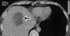
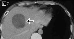
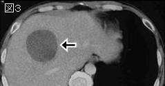
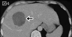
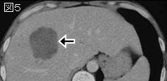
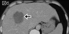
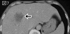
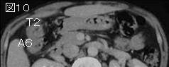
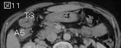
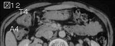
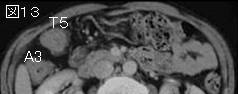
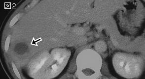
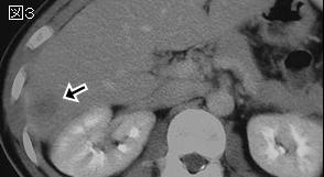
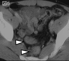
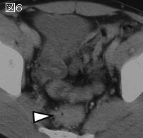
要旨:感染者便中の赤痢アメーバ(Entamoeba histolytica)成熟嚢子(cyst)が,経口感染から腸管感染をきたし,大腸で増殖し栄養型(trophozoite)となって経門脈的に肝臓に達し膿瘍形成したものである.腸管型アメーバ症の慢性期に発症し腸管症状はみられないことも多い.世界的には熱帯と亜熱帯地域に多い.日本では近年増加傾向にあり,輸入感染症,同性愛者の感染,免疫不全者の発症が注目されている.12:1と圧倒的に男性に多く,30〜50歳代に好発する.糞便や膿汁から赤痢アメーバ原虫を検出すれば確診となるが,検出率は11〜30%と低い.間接赤血球凝集反応(IHA)は100%の確信率を有する.薬物治療はmetronidazoleが第一選択で治癒率は90%以上である.ドレナージの適応は,1:薬物療法によって症状の改善がみられない,膿瘍が縮小しない,2:膿瘍の破裂・穿破のおそれがあるもの,特に心外膜への波及のおそれのある左葉の病変,3:二次感染が疑われる症例.
参考文献
Radiographics. 2001 Jul-Aug;21(4):895-910.
Cystic focal liver lesions in the adult: differential CT and MR imaging features.
Mortele KJ, Ros PR.
Cystic lesions of the liver in the adult can be classified as developmental, neoplastic, inflammatory, or miscellaneous. Although in some cases it is difficult to distinguish these entities with imaging criteria alone, certain cystic focal liver lesions have classic computed tomographic (CT) and magnetic resonance (MR) imaging features, which are important for the radiologist to understand and recognize. Lesions with such features include simple (bile duct) cyst, autosomal dominant polycystic liver disease, biliary hamartoma, Caroli disease, undifferentiated (embryonal) sarcoma, biliary cystadenoma and cystadenocarcinoma, cystic subtypes of primary liver neoplasms, cystic metastases, pyogenic and amebic abscesses, intrahepatic hydatid cyst, extrapancreatic pseudocyst, and intrahepatic hematoma and biloma. Specific CT and MR imaging findings that are important to recognize are the size of the lesion; the presence and thickness of a wall; the presence of septa, calcifications, or internal nodules; the enhancement pattern; the MR cholangiographic appearance; and the signal intensity spectrum. In addition, access to critical clinical information remains extremely important. The most important clinical parameters defined include age and gender, clinical history, and symptoms. An understanding of the classic CT and MR imaging appearances of cystic focal liver lesions will allow more definitive diagnosis and shorten the diagnostic work-up. PMID: 11452064
|