|
文献考察:瘻(孔).fistula:一つの上皮で覆われている表面から他の上皮で覆われている表面への異常な導管.
Radiology. 2002 Jul;224(1):9-23.
Acquired gastrointestinal fistulas: classification, etiologies, and imaging evaluation.
Pickhardt PJ, Bhalla S, Balfe DM.
Fistulas are abnormal communications between two epithelial-lined surfaces. Gastrointestinal fistulas encompass all such connections that involve the alimentary tract, and they can be congenital or acquired in nature. This review focuses on acquired gastrointestinal fistulas. Development of an acquired gastrointestinal fistula can greatly affect patient outcome, yet the clinical manifestations are often protean in nature and the etiology, elusive. Imaging plays an important role in the detection and management of acquired gastrointestinal fistulas. The more routine use of cross-sectional imaging (especially computed tomography and magnetic resonance imaging) has altered the standard sequence of radiologic evaluation for possible fistulas, but fluoroscopic studies remain a valuable complement, especially for confirming and defining the anomalous communications. In this review, a classification scheme for gastrointestinal fistulas is provided, major causes are discussed, and individual fistula types are elaborated with an emphasis on contemporary imaging approaches. PMID: 12091657
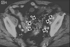
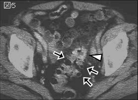
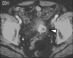
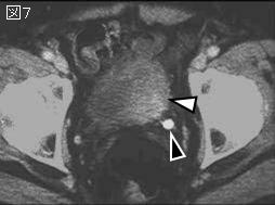
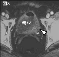
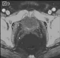
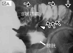
追記:分類は表1,原因は表2.腸管膀胱瘻の原因で最も多いのはS状結腸憩室炎(S状結腸膀胱瘻),次いでCrohn病(小腸膀胱瘻),さらにS状結腸または直腸癌や膀胱癌である.
|


;)
;)