その他(Miscellaneous)シリーズ6 EXPERT COURSE 解答 【症例 ME 26】

原発性腹部大動脈十二指腸瘻.Primary aortoduodenal fistula


吐血例で胃内の※は血腫を意味し,横行結腸(T)と上行結腸(A)の液状内容物は血液または血腫を示唆する.図8〜図16に,最大径5〜6cmの腹部大動脈瘤がある(↑)が,それは下血の原因か? 図9〜図13の,瘤内血腫内の造影剤(△)は血液が流れている真腔から瘤内の古い血腫内へ解離を起こしている現象である.はっきりしたextravasationは指摘できないが,Treitz靱帯直下の空腸図7の1〜図11の20までの上部空腸は,他の小腸と比較して高吸収値を呈しており,腸管内へ造影剤の漏出を反映している可能性が高い(early phaseとdelayed phaseのdouble phaseの造影CTを撮るとextravasationがはっきりすると思われる).図8と図9で動脈瘤の頭側先端と十二指腸水平部が交差しているので,そこで大動脈十二指腸瘻(aortoduodenal fistula)を形成して,腸管内穿破していると推測する.Du:十二指腸.内視鏡検査で胃と十二指腸内に大量の凝血塊を認め,食道胃接合部に露出血管を伴うマロリー・ワイス症候群を認めるが活動性の出血はない.大学病院へ転送し緊急手術が施行され,腹部大動脈十二指腸瘻が確認された.


参考文献
J Comput Assist Tomogr. 2004 May-Jun;28(3):343-7.
Multidetector computed tomography imaging of aortoenteric fistula.
Perks FJ, Gillespie I, Patel D.

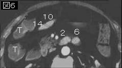
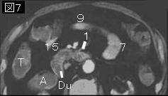
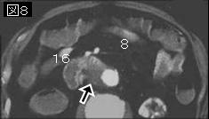
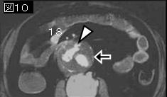
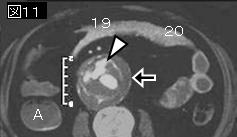
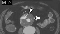
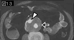
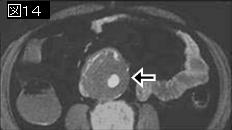
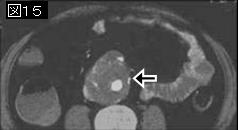
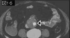
Three case reports illustrating multidetector computed tomography (CT) imaging findings of secondary aortoenteric fistula (AEF) are described and presented in axial sections, multiplanar reformats, and 3-dimensional reconstruction. Fistulae occurred in the early and late postgrafting period and involved both end-to-end and end-to-side aortic graft anastomoses. Multidetector CT is quick and accurate in the diagnosis of bleeding AEF. PMID: 15100538

 【 次の問題→ 】  【 このシリーズの問題一覧に戻る 】 【 演習問題一覧に戻る 】