|
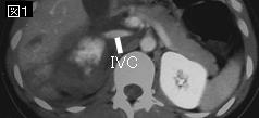
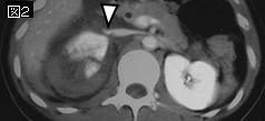
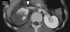
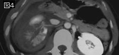
文献考察:小児の鈍的腎外傷のCT検査の適応
1)Urol Clin North Am. 2006 Feb;33(1):33-40
The diagnosis, management, and outcomes of pediatric renal injuries.
Buckley JC, McAninch JW.
Most pediatric renal trauma is minor and poses no significant danger to the child. A small percentage of children sustain a severe renal injury that demands immediate evaluation and decision of operative versus nonoperative management. Selective management of pediatric renal trauma based on mechanism of injury, hemodynamic stability,associated nonrenal injuries, and CT imaging has led to a renal exploration rate of 5%to 11% with renal salvage rates of more than 98%.PMID: 16488278
要旨:循環動態が安定しており,血尿または尿沈渣で赤血球:>50/hpf,転落や交通事故などの急な減速による外傷はCT検査(表1).
2)J Urol. 2002 Jun;167(6):2543-6; discussion 2546-7.
Blunt traumatic hematuria in children. Is a simplified algorithm justified?
Perez-Brayfield MR, Gatti JM, Smith EA, Broecker B, Massad C, Scherz H, Kirsch AJ.
PURPOSE: We determined whether radiographic evaluation is indicated in all children with traumatic hematuria. MATERIALS AND METHODS: We retrospectively reviewed the records of 110 children from 1992 to 1999 diagnosed with blunt trauma and hematuria. It is routine practice at our emergency department to perform radiographic evaluation in all children with hematuria regardless of the degree. Each chart was evaluated for the mechanism of injury, degree of hematuria, hypotension, imaging studies, renal injury, renal anomalies, associated injuries and outcome. RESULTS: A total of 110 patients 1 to 18 years old (mean age 9) were identified. The most common mechanism of injury was motor vehicle accident in 37 children (34%), followed by a fall in 32 (29%). Grades I to V renal injury was present in 5, 6, 6, 6 and 1 cases, respectively (22%), while 1 (0.9%) involved ureteropelvic junction avulsion. No child had renal pedicle injury. In 9 patients renal anomalies were detected incidentally. Of the 110 patients 101 underwent radiographic evaluation, including computerized tomography in 97 (88%). The 24 patients (22%) with significant renal injury and all with incidentally diagnosed renal anomalies had 50 or greater red blood cells per high power field on urinalysis, while 1 with ureteropelvic junction avulsion presented without hematuria. Hypotension was present in only 3 patients (2.7%), who also had associated injuries, including 2 who presented with renal injury. All 3 with associated injuries. Associated injuries were identified in 11 of 25 patients (44%). The 9 patients (8%) who did not undergo radiographic imaging had negative results on repeat urinalysis with an excellent outcome. CONCLUSIONS: We recommend that radiological evaluation consisting of abdominal and pelvic computerized tomography should be performed only in patients with 50 or greater red blood cells on urinalysis, hypotension at presentation to the emergency room or based on the severity of mechanism of injury, for example high speed motor vehicle accident deceleration injuries. The patient who presented with ureteropelvic junction avulsion without hematuria would have undergone imaging considering the mechanism of injury and number of associated injuries.PMID: 11992085
要旨:上記文献と同様なCT検査の適応(表2).
|


;)
;)