|
文献考察:外傷性横隔膜へルニア
1)【ヘルニアの画像診断】 胸部・横隔膜のヘルニア(解説/特集)
Author:金井信恭(聖マリアンナ医科大学 放射線医学), 滝澤謙治, 森田あかね, 八木橋国博, 新美浩, 栗原泰之, 中島康雄
Source:臨床放射線(0009-9252)48巻6号 Page711-717(2003.06)
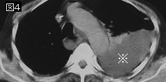
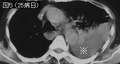
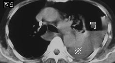
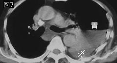
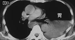
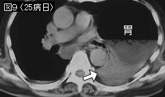
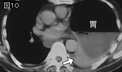
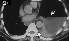
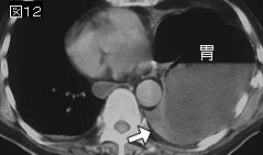
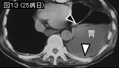
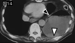
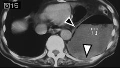
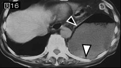
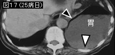
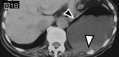
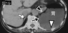
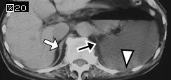
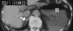
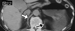
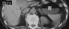
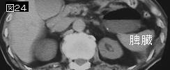
要旨:多くは鈍的腹部外傷に合併する.80〜90%は左側に発生し,ヘルニア臓器は大網,胃,結腸,脾臓が多い.腹腔内臓器が胸腔内へ脱出すると患側肺の虚脱や縦隔の偏位をきたし呼吸循環障害を起こすことがある.CTで有用な所見は,1:dependent viscera sign(肝上1/3が右後方肋骨に,腸や胃が左後方肋骨に接する),2:collar sign(横隔膜断裂部による消化管のくびれ),3:subhepatic hemothorax(胸腔内に脱出した肝が背側胸壁に密着し血胸を上下に二分する)などがいわれている.マルチスライスCTのmultiplanar reformation:MPR像(多断面再構築法)は横隔膜の非連続性(断裂)が直接確認可能である.
2)外傷性横隔膜へルニアのCT所見
AJR Am J Roentgenol. 2005 Jan;184(1):24-30.
Helical CT of blunt diaphragmatic rupture.
Nchimi A, Szapiro D, Ghaye B, Willems V, Khamis J, Haquet L, Noukoua C, Dondelinger RF.
OBJECTIVE: This study evaluated CT findings for signs of blunt diaphragmatic rupture. MATERIALS AND METHODS: CT examinations of 179 blunt trauma patients, including 11 with left-sided and five with right-sided blunt diaphragmatic rupture, were reviewed by two staff radiologists who first decided by consensus on the presence or absence of 11 published signs of blunt diaphragmatic rupture and then formulated the diagnosis in terms of absence of, presence of, or suggestion of blunt diaphragmatic rupture. The significance of the findings was assessed by multivariate logistic regression. Four other reviewers interpreted the CT findings independently. They were asked first to formulate a diagnosis in terms of absence of, presence of, or suggestion of blunt diaphragmatic rupture and then to enumerate the findings supporting a diagnosis or suggestion of blunt diaphragmatic rupture. These findings were compared with those of the staff radiologists. RESULTS: Diaphragmatic discontinuity, diaphragmatic thickening, segmental nonrecognition of the diaphragm, intrathoracic herniation of abdominal viscera, elevation of the diaphragm, and both hemothorax and hemoperitoneum were strong predictors of blunt diaphragmatic rupture (p
要旨:外傷性横隔膜へルニアの11CT所見中高率に認めるのは,1:横隔膜の断裂(discontinuity),2:横隔膜の肥厚(curled diaphragm sign:10mm以上),3:横隔膜の部分的消失(segmental nonrecognition of diaphragm),4:腹腔内臓器の胸腔内脱出,5:横隔膜挙上(右側は左横隔膜の頂上より5cm以上,左側は右横隔膜頂上より4cm以上の部位で腹腔内臓器を認める),6:血胸と腹腔内血液の存在.その他の5つの所見とは,1:collar sign(横隔膜破裂部で消化管が締め付けられてwaistlike appearanceを呈する),2;dependent viscera sign(右側では肝臓上1/3が右後方肋骨に,左側では腸管や胃が左後方肋骨に接する:下記文献参照),3:胸腔内貯留液が腹腔内臓器に接する(ヘルニアがなければ両者の間に肺が介在する).4:横隔膜断裂部からの出血を示す横隔膜の位置で造影剤の漏出(extravasation)を認める.5:肋骨骨折.
3)AJR Am J Roentgenol. 2001 Nov;177(5):1137-40.
The "dependent viscera" sign in CT diagnosis of blunt traumatic diaphragmatic rupture.
Bergin D, Ennis R, Keogh C, Fenlon HM, Murray JG.
OBJECTIVE: The objective of our study was to describe the "dependent viscera" sign and determine its usefulness at CT in the diagnosis of diaphragmatic rupture after blunt abdominal trauma. MATERIALS AND METHODS: The study sample consisted of 28 consecutive patients (19 men, nine women) between 17 and 74 years old (mean age, 31 years) who had undergone abdominal CT and subsequent emergency laparotomy after a blunt trauma. Ten patients had a diaphragmatic rupture (six, right-sided; four, left-sided) at laparotomy. An experienced radiologist unaware of the surgical findings retrospectively reviewed the CT scans, and then a second radiologist reviewed the scans to provide interobserver agreement. Note was made of discontinuity of the diaphragm, intrathoracic herniation of abdominal contents, and waistlike constriction of bowel (the collar sign). Also noted was whether the upper one third of the liver abutted the posterior right ribs or whether the bowel or stomach lay in contact with the posterior left ribs. Either of these findings was termed the "dependent viscera" sign. The radiologists' detection rate of diaphragmatic rupture on the CT scans via observance of the dependent viscera sign was determined. Interobserver agreement was assessed using Cohen's kappa statistic. RESULTS: The dependent viscera sign was observed on the CT scans of 100% of the patients with a left-sided diaphragmatic rupture and of 83% of the patients with right-sided diaphragmatic rupture. Both observers missed one case of right-sided diaphragmatic rupture. The radiologists' overall rate of detecting diaphragmatic rupture was 90% using the dependent viscera sign. We found excellent interobserver agreement (kappa = 1) for detection of the dependent viscera sign and for the diagnosis of diaphragmatic tear on CT scans. CONCLUSION: The dependent viscera sign increases the detection at CT of acute diaphragmatic rupture after blunt trauma.PMID: 11641188(full text)
追記:横隔膜断裂がないと腹腔内臓器は横隔膜に支持され,後方肋骨との間に肺が介在する.断裂があり腹腔内臓器が脱出すると横隔膜の支持を失い腹腔内臓器は後方に肋骨まで落下する.従って,右側では肝臓の上1/3,左側では胃と他の消化管が後方肋骨に接する所見は横隔膜へルニアを示唆する.
|


;)
;)
;)
;)