|
文献考察:結核性腹膜炎
1)結核性腹膜炎,乾性型結核性腹膜炎
Author:星進悦(釜石市民病院)
Source:日本臨床(0047-1852)別冊腹膜・後腹膜・腸間膜・大網・小網・横隔膜症候 Page22-25(1996.05)
2)検査値の読み方 腹水中adenosine deaminase(ADA)高値が診断に有用であった結核性腹膜炎の1例
Author:花尻和幸(東京逓信病院 消化器科), 橋本直明, 高倉裕一, 小林克也, 関川憲一郎, 松川雅也, 松浦広, 鈴木丈夫, 是永建雄
Source:臨床消化器内科(0911-601X)18巻2号 Page253-258(2003.01)
Abstract:27歳男.1ヵ月続く腹痛,盗汗,体重減少を主訴に受診した.腹部所見,炎症反応高値より,腹膜炎を疑い,即日入院した.画像所見及び腹水中adenosine deaminase(ADA)活性の上昇を認めたため,結核性腹膜炎と診断した.第10病日より抗結核薬(isoniazid,rifampicin,ethambutol)の投与を開始した.治療により解熱し,盗汗,腹痛の消失,CRPの陰性化,血沈の正常化を認めた.腹部,骨盤部CT上,腹水の消失を確認し,第71病日に退院した.退院直前に大腸内視鏡と注腸造影を行ったが,腸結核を疑わせる所見は認められなかった.第10病日に採取した胃液培養で結核菌が2コロニーと陽性であったことが,第36病日に判明した.
3)Abdom Imaging. 2001 May-Jun;26(3):319-22.
Tuberculous peritonitis: a diagnostic challenge.
Hiller N, Lioubashevsky N.
Tuberculous (TB) peritonitis rarely occurs in developed countries. The clinical presentation and laboratory tests are usually insufficient for diagnosing TB peritonitis and distinguishing it from peritoneal carcinomatosis. We present two cases of TB peritonitis and illustrate the computed tomographic findings of the disease according to the world literature. Recognition of the radiologic manifestations and maintenance of a high index of suspicion in population at risk have paramount importance for diagnosing this uncommon disease.PMID: 11429963
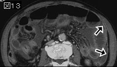
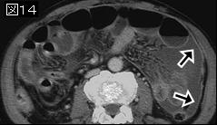
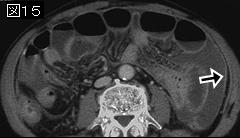
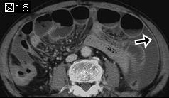
追記:肉眼的特徴により3型に分類される
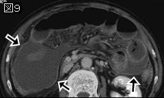
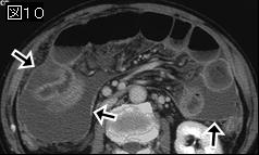
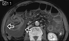
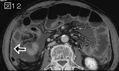
Three, usually overlapping, forms of the CT appearance of TB peritonitis have been described.
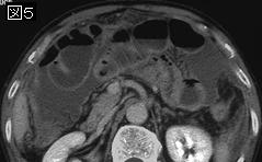
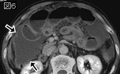
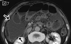
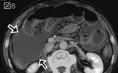
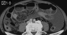
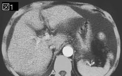
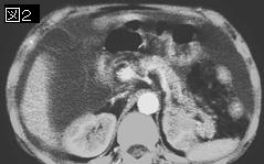
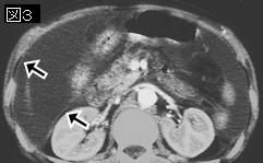
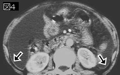
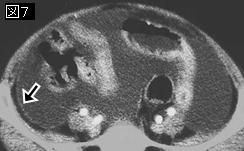
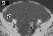
1. The "wet ascitic" type occurs in 90% of cases and is characterized by large viscous ascites diffusely distributed or loculated into complex pockets.On CT, the fluid had high attenuation values (25-45 HU) secondary to the high protein and cellular content.
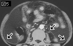
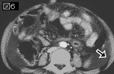
2. The "fibrotic fixed" type is seen in 60% of cases and is characterized by large mass formation of the omentum, adhesions of mesentry, matting of bowel loops and mesentery, and, occasionally, loculated ascites. The CT presentation of that type of peritonitisis of low-density masses and nodular soft tissue along the hypervascular peritoneal surfaces, mesentery, and omentum. Tethering of bowel loops and stellate appearance of bowel loops may also be observed.
3. The ”dry" type of peritonitis is seen in 10% of cases and is characterized by fibrous peritoneal reaction, enlarged caseous lymph nodes, and dense adhesions.
4)Am J Surg. 2003 Jun;185(6):567-73.
Indication for peritoneal biopsy in tuberculous peritonitis.
Chow KM, Chow VC, Szeto CC.
BACKGROUND: With the introduction of effective antituberculous chemotherapy, the clinical outcome of tuberculous peritonitis depends much on the diagnostic accuracy of this disease entity. This review summarizes the current state-of-the-art thinking regarding the protean manifestation and diagnostic modalities of this major infectious disease. DATA SOURCES: This review was compiled after an extensive search of the current and historical literature, comprising 1,070 cases of tuberculous peritonitis. A number of important areas were highlighted, with emphasis on the diagnostic value and clinical impact of peritoneal biopsy. CONCLUSIONS: We believe an aggressive diagnostic approach, particularly with peritoneal biopsy, is warranted for the diagnosis and timely treatment of tuberculous peritonitis.PMID: 12781888
4文献の要約:結核性腹膜炎は3型に分類される. 1)大量の腹水を伴い,腹膜に粟粒結節を認める滲出型,2)大網腫瘤,腸管・腹膜の線維性癒着が著明で,浸出液の少ない癒着型,3)腹膜の線維化・癒着,結核腫の乾酪化の著明な乾酪型.
結核性腹膜炎は全結核症の0.04-0.55%とまれな疾患であり, 特徴的な所見に乏しく, 癌性腹膜炎との鑑別も困難なことから, 診断や治療の遅延あるいは困難な例の存在が指摘されている.
症状は病型や病期によってさまざまであり, 特有なものはない. 一般に慢性に経過し, 発熱, 全身倦怠感,下痢, 腹痛, 腹部膨満感などが主症状である.
腹水所見は本症の診断上重要である.腹水中のADA(adenosine deaminase)とCA125が有意に高値を示し,マーカーとして診断, 治療, 経過観察に有用であることが報告されている. また, 本症の腹水は蛋白濃度が2.5g/dl以上の滲出液で, 細胞診では成熟リンパ球が主体を占める.
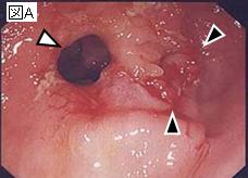
本症の診断は, 結核菌を証明するか, 結核に特有な乾酪性肉芽腫を組織的に証明するかのいずれかである. しかしながら, 腹水中の結核菌が検出される率は, 10%前後と低率である. しかも, 結核菌は増殖が遅いため小川培地では検出までに4-6週間を要する. このため最近では,迅速な菌検出方法としてDNAプローブ法が注目されている.さらに, PCR(polymerase chain reaction)法を適用して数時間で検体中の結核菌の存在を証明することができるようになってきている. 細菌学的診断が困難な場合には, 開腹生検, 手術時生検, 腹腔鏡下生検などの病巣部の組織学的検索による確定診断が有力となる.
|