CT of Acute Abdomen

Epigastric Pain series 3 RESIDENT COURSE Answer [ER Case 15]

Perforated gastric cancer


There is ascites (reference mark) in the left subphlenic space (left side of stomach) in Fig.1-Fig.6, and white arrowhead of Fig.6-Fig.8 is free air. Because circled number 12 of Fig.5-Fig.11 indicate duodenum, wall thickening (black arrows) of Fig.6-Fig.10 is a lesion of antrum. It is circumferential, and irregular enhancement by contrast strongly suggests an advanced malignant tumor. A black arrowhead of Fig.9 is very likely to be an enlarged lymph node. Perforation of giant Borr 3 type gastric cancer (Fig.A: black arrow) of antrum with metastases to lymph nodes at the lesser curvature side was confirmed by surgery. There was no metastatic lesion in liver indicating white arrows of Fig.9 are small cysts.


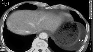
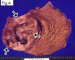
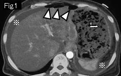
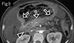
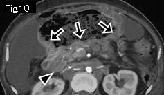
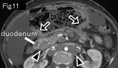
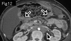
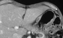
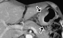
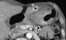
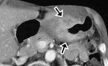
Reference case 1 (5mm slice, gastric cancer perforation): A 53-year-old female with schizophrenia. Over one month, she developed appetite loss, anemia, and gradually distending abdomen. She presented with increasing epigastric pain for several hours. Temperature: 37.5 degrees Celsius, abdomen showed signs of generalized peritonitis.
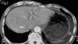
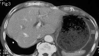
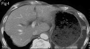
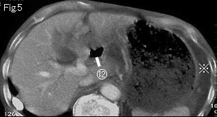
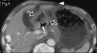
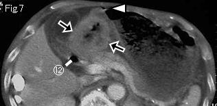
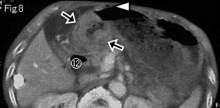
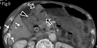
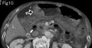
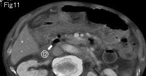
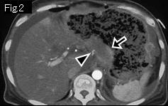
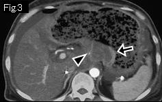
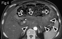
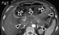
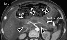
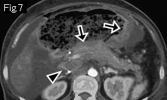
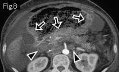
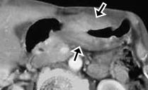
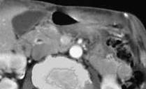
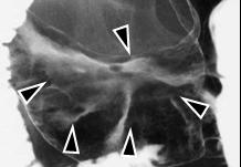
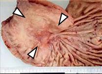
Ascites (reference mark) and free air (white arrowheads) in Fig.1 indicate gastrointestinal perforation. Stomach is dilated with food residue. Black arrows in Fig.2-Fig.12 show contrast-enhanced wall thickening in comparison with the other part, and black arrowheads are enlarged lymph nodes, which leads to a diagnosis as perforation of advanced gastric cancer. Surgery and pathological examination revealed perforation of gastric cancer with extensive invasion into retroperitoneum.


Reference case 2 (non-perforated gastric cancer): A 56-year-old male presented with loss of appetite and vomiting.
Black arrow, black arrowhead and white arrowhead manifest advanced gastric cancer that extends from gastric angle to antrum.


 Previous Case   to Questions Page   to Practical Exercises Page