CT of Acute Abdomen

Epigastric Pain series 3 EXPERT COURSE Answer [EE Case 15]

Penetrated gastric ulcer


There is no free air or ascites. Thickening by submucosal edema (black arrowhead) begins from Fig.8, and extends to posterior wall of antrum of Fig.13. White arrow of Fig.11 is intragastric gas. On the other hand, gas (black arrow) of Fig.11-Fig.13 should be considered to be a wall defect (namely ulcerative lesion) of posterior wall because of associated wall thickening by submucosal edema. Fig.14 and Fig.15 show fat stranding (white arrowheads) at caudal site of ulcerative lesion suggesting gastric ulcer penetration. Surgery revealed gastric ulcer of posterior wall penetrated into transverse colon mesentery. Fig.A shows serosal side of resected specimen and black arrowhead is penetration site, Fig.B shows mucosal side and black arrow is an ulcer.


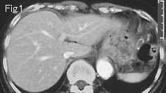
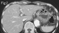
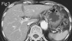
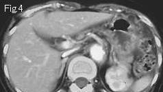
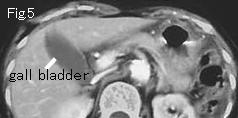
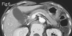
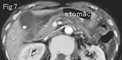
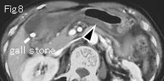
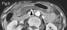
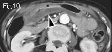
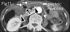
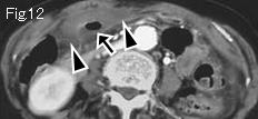
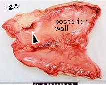
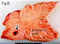
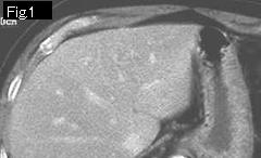
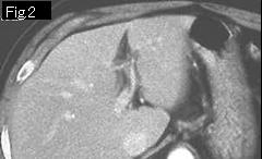
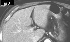
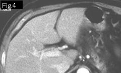
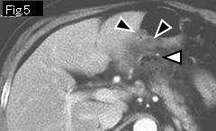
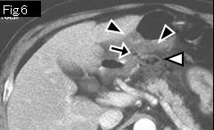
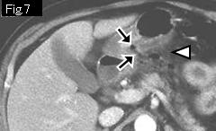
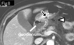
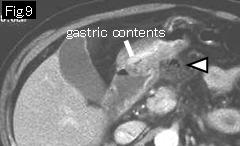
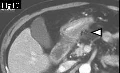
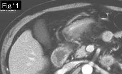
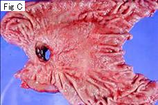
Reference case: (gastric ulcer perforation): A 57-year-old male with a past history of duodenal ulcer 9 years ago, presented with gradually aggravating epigastric pain for 24 hours. Temperature: 36.7 degrees Celsius. There is tenderness in epigastrium. No free air is seen directly beneath the anterior abdominal wall. In Fig.1-Fig.4, gastric wall does not show submucosal edema to indicate an acute lesion. Pyloric wall manifests submucosal edema from Fig.5 (black arrowhead), and Fig.6-Fig.8 show gas and defect (black arrow) in the posterior wall that is thickened by submucosal edema. Note that free air and food debris (white arrowhead) in omental sac of Fig.5-Fig.10 are depicted. As a result, diagnosis as perforation of ulcerative lesion of pyloric posterior wall of stomach can be made. Fig. C is resected specimen and pathology reported as benign gastric ulcer with perforation.


 Previous Case   to Questions Page   to Practical Exercises Page