|
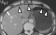
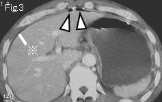
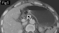
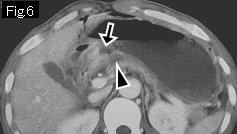
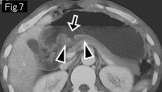
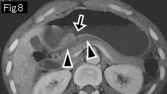
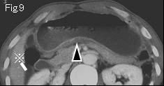
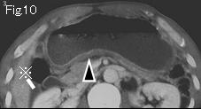
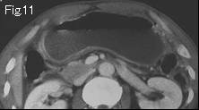
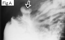
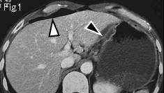
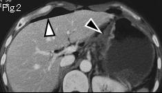
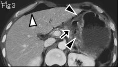
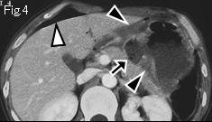
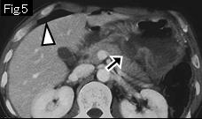
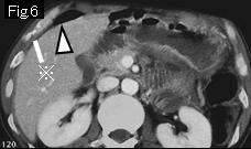
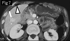
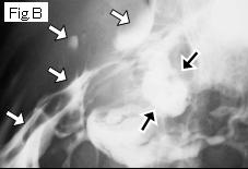
Reference case (perforated gastric ulcer): A 43-year-old male presented with sudden onset of intense epigastric pain for three hours. Temperature: 36.4 degrees Celsius. Physical examination revealed clinical signs of peritonitis from epigastrium to umbilical area.CT images show free air (white arrowhead) and a little ascites (reference mark). The lesser curvature of stomach manifests edematous wall thickening (black arrowheads), and black arrow is an acute ulcer that spreads from gastric angle through posterior wall. Gastrografin contrast study (Fig.B) showed an ulcer (black arrow) in posterior wall and massive leakage (white arrow) of contrast agent into free peritoneal cavity. In spite of the fact that leakage of contrast medium into free peritoneal cavity mean definite indication of surgery, conservative treatment for two weeks succeeded for this case, which should be considered as exceptional.
|