|
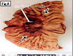
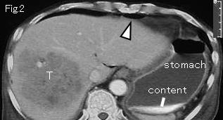
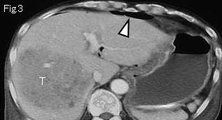
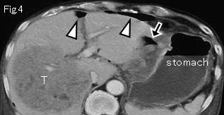
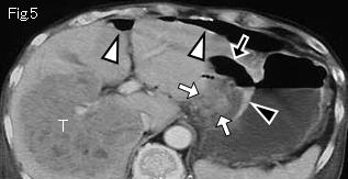
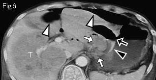
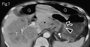
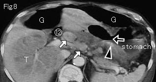
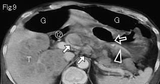
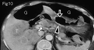
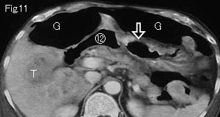
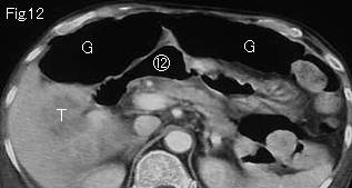
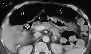
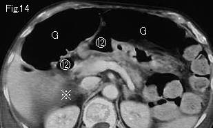
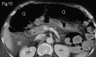
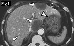
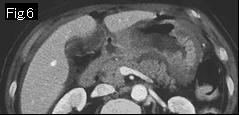
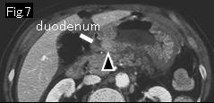
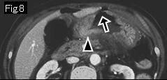
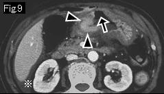
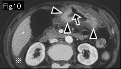
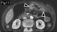
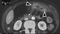
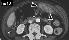
All images show massive free air (white arrowheads and G), and there is ascites (reference mark) in the Morison pouch in Fig.13-Fig.15. As for the large lesion (T) of liver of Fig.2-Fig.12, irregular internal structure is highly suggestive of metastatic tumor. Black arrows of Fig.4-Fig.11 indicates a deep ulcerative lesion, and mucosa of stomach (black arrowheads) of Fig.5-Fig.10 is slightly thickened and strongly contrast-enhanced comparing with the other part, suggesting a neoplastic lesion. White arrows of Fig.5-Fig.10 are very likely to be enlarged lymph nodes. Therefore, perforated gastric cancer with metastases to liver is a diagnosis. Circled number 12: duodenum. Perforation of gastric cancer (Fig.A: black arrows) with liver and lymph node metastasis was confirmed by surgery and pathology.
|
|
Reference case 1 (5mm slice, gastric cancer perforation): A 70-year-old female. She had occasional epigastric pain for a few days, which suddenly increased in intensity nine hours ago. Temperature: 37.5 degrees Celsius, physical examination revealed tenderness and rebound tenderness in epigastrium, but without muscle guarding.
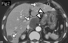
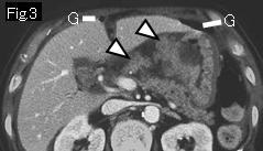
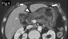
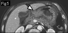
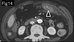
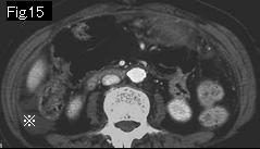
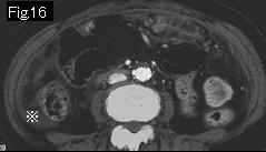
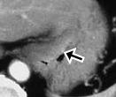
Free air (G) of Fig.1-Fig.4 and ascites (reference mark) of Fig.9-Fig.16 strongly indicate gastrointestinal perforation. The hepatic lesion (white arrows) of Fig.1 and Fig.2, and enlarged lymph nodes (white arrowhead) of Fig.1-Fig.5 suggest metastasis of malignant tumor. Black arrowheads of Fig.8-Fig.14 show wall thickening with irregular contrast enhancement indicating gastric cancer, and black arrow of Fig.8-Fig.10 is an ulcer leading to a diagnosis as perforation of gastric cancer. Surgery revealed a perforated firm tumor of 5cm size in anterior wall of stomach and several metastatic nodes in lesser curvature side as well as liver metastasis. Pathology reported as gastric cancer.
|
|
Reference case 2(gastric cancer): A 54-year-old female with epigastric pain for 24 hours.
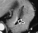
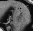
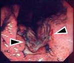
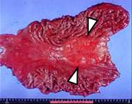
Black arrow of CT images, black arrowheads of endoscopy and white arrowheads of resected specimen show gastric cancer. Pathology: adenocarcinoma.
|
|