|
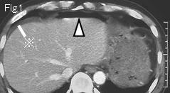
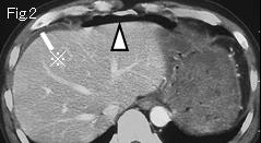
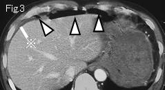
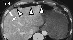
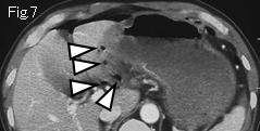
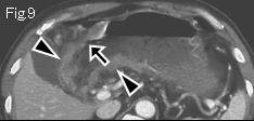
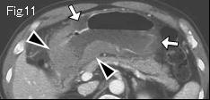
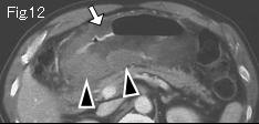
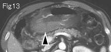
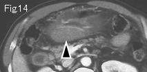
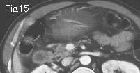
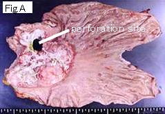
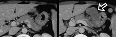
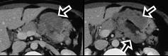
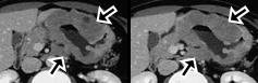
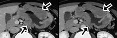
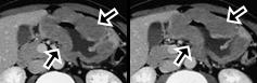
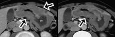
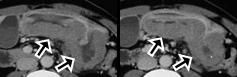
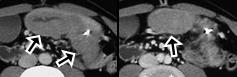
There is free air (white arrowheads) in Fig.1-Fig.4 and ascites (reference mark) around liver. White arrow of Fig.11 and Fig.12 presents water density consistent with submucosal edema, on the other hand, wall thickening (black arrowheads) of antrum of Fig.9-Fig.14 demonstrates contrast enhancement suggesting high possibility of neoplastic lesion. No symptoms until two days ago in spite of circumferential and extensive spread in antrum indicates that a lesion has considerable degree of distensibility suggesting malignant lymphoma. Adjacent free air (white arrowheads) in Fig.7 suggests that black arrow of Fig.9 and Fig.10 is a perforation site. Subtotal gastrectomy was undertaken (Fig.A) and pathological examination reported as malignant lymphoma (large cell type).
|