|
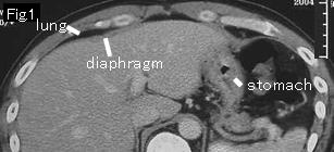
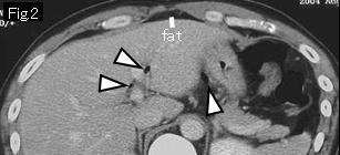
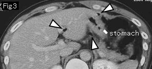
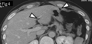
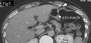
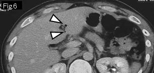
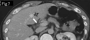
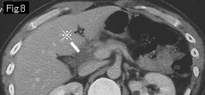
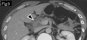
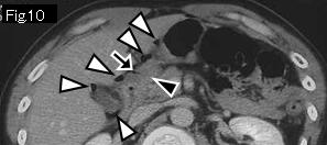
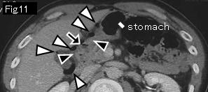
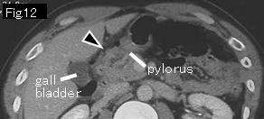
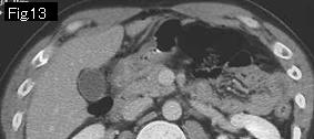
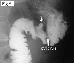
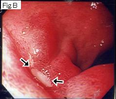
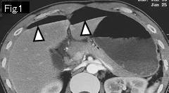
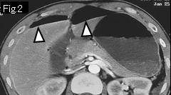
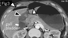
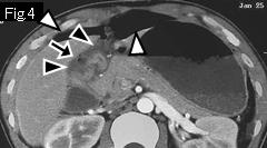
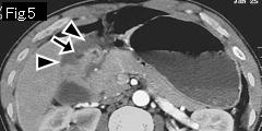
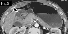
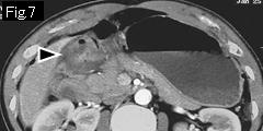
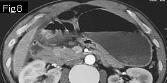
All white arrowheads in Fig.2-Fig.6 are free air. Because of no submucosal edema in gastric wall, there seems to be no acute lesion in stomach. Anterior wall of duodenal bulb demonstrates submucosal edema (black arrowheads) in Fig.9-Fig.12, and black arrow of Fig.10 and Fig.11 indicates an acute ulcerative lesion. There is extraluminal gas (white arrowheads) around the duodenum, and ascites in Fig.7 and Fig.8 (reference mark), which leads to the diagnosis as duodenal ulcer perforation. Contrast study by gastrografin was undertaken for confirmation. White arrow of Fig.A indicates duodenal ulcer crater. Because no leakage of contrast agent was demonstrated, conservative treatment was chosen with success. Endoscopy on the tenth day showed an ulcer in anterior wall of duodenal bulb (Fig.B: black arrows).
|