|
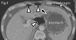
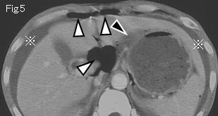
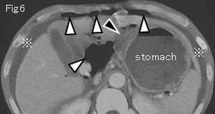
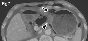
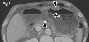
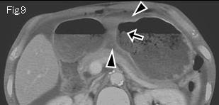
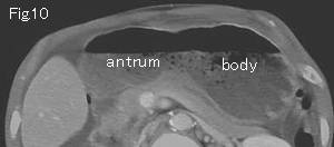
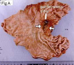
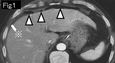
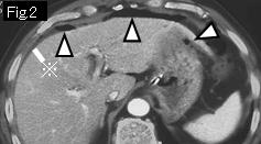
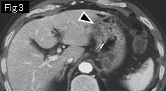
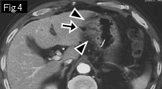
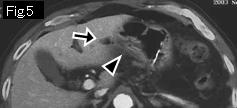
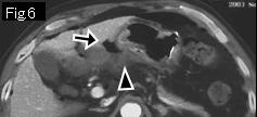
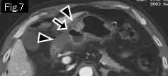
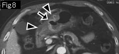
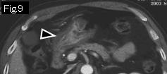
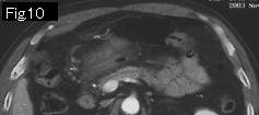
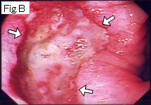
Reference case (perforated gastric ulcer): A 61-year-old male with a past history of gastric ulcer one year ago, presented with sudden onset of intense epigastric pain which occurred two hours ago. Temperature: 36.1 degrees Celsius. Physical examination showed tenderness, rebound tenderness and muscle guarding in epigastrium. There are free air (white arrowhead) and ascites (reference mark) in Fig.1 and Fig.2, and black arrowheads of Fig.3-Fig.9 indicate wall thickening by submucosal edema of antrum, and gas (black arrow) of Fig.4-Fig.8 is an acute ulcerative lesion. Responding to conservative treatment, abdominal pain was relieved on the next day. Endoscopy performed eight days later revealed a giant ulcer in antrum (Fig.B: white arrows).
|