CT of Acute Abdomen

Epigastric Pain series 2 EXPERT COURSE Answer [EE Case 8]

Perforated gastric ulcer of posterior wall


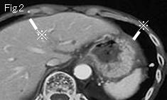
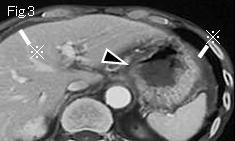
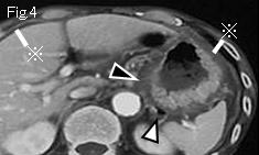
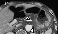
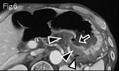
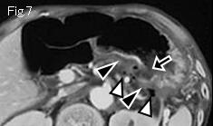
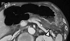
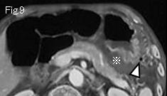
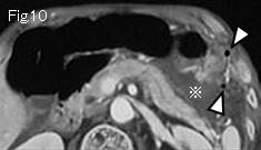
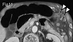
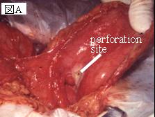
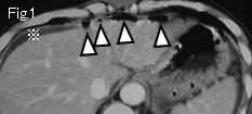
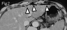
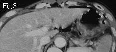
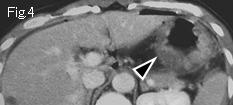
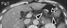
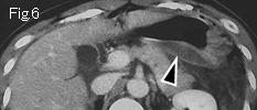
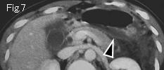
There is ascites (reference mark) in dorsal site of stomach of Fig.9-Fig.11, as well as around liver and stomach of Fig.2-Fig.4. White arrowheads of Fig.4-Fig.11 indicate extraluminal free air that suggest perforation of posterior wall of stomach. Edematous thickening begins with the lesser curvature of stomach of Fig.3 (black arrowhead), and extends to posterior wall of stomach of Fig.7. Black arrow of Fig.6 and Fig.7 is a wall defect, and diagnosis as perforation of posterior wall ulcer of stomach can be made. Fig.A is perioperative photograph which confirmed above findings.


Reference case (perforated gastric ulcer): A 47-year-old female had occasional epigastric pain for two weeks. Pain became intense several hours ago. Temperature: 38.3 degrees Celsius. Examination revealed tenderness, rebound tenderness and muscle guarding in the whole abdomen. Fig.1 and Fig.2 show free air (white arrowheads) and ascites (reference mark). Edematous wall thickening (black arrowheads) of posterior wall of stomach is seen in Fig.4 - Fig.7, and black arrow of Fig.5 indicate an acute ulcerative lesion that is highly suggestive of perforated posterior wall ulcer. Contrast study showed leakage (white arrows of Fig.B ) of contrast medium. The findings were confirmed at surgery.


 Previous Case    Next Case   to Questions Page   to Practical Exercises Page