CT of Acute Abdomen

Epigastric Pain series 1 RESIDENT COURSE Answer [ER Case 4]

Perforated gastric ulcer


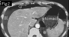
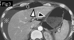
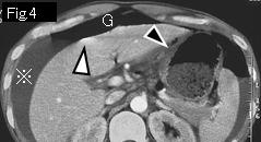
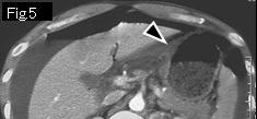
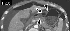
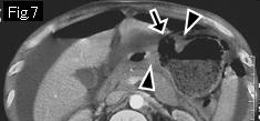
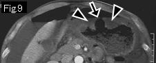
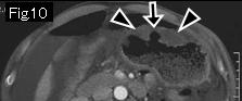
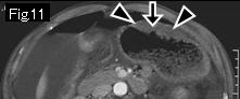
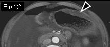
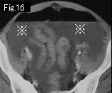
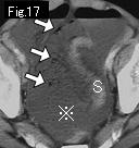
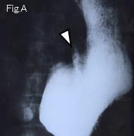
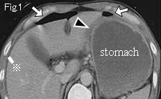
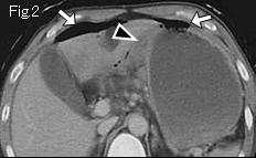
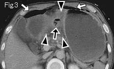
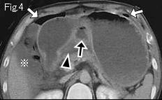
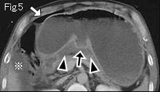
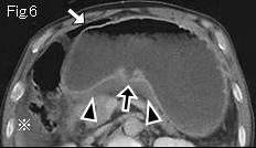
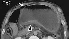
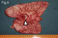
Presence of massive ascitic fluid (reference mark), free air (G), edematous wall thickening at lesser curvature of stomach (Fig.3: black arrowheads), and an ulcerative lesion extending from gastric angle to anterior wall of stomach (Fig.6–Fig.11: black arrows) are indicative of gastric ulcer perforation. Foamy gas (white arrows) of Fig.17 should be considered as food debris (leaked intestinal content). S: sigmoid colon. Upper GI series shows a giant gastric ulcer (Fig.A: white arrowhead). Laparotomy revealed perforated gastric ulcer(2cm in size) at anterior wall of stomach.


Reference Case (gastric ulcer perforation): A 46-year-old male had occasional epigastric pain since a duodenal ulcer disease was diagnosed ten years ago. He had been taking commercially available anti-ulcer drugs. The patient had had tarry stools for two weeks and sudden onset of severe epigastric pain for five hours.
CT images show ascitic fluid (reference mark) and free air (white arrows). Edematous gastric wall thickening extends to posterior wall of stomach(Fig.1-Fig.7, black arrowheads). The ulcerative lesion extends from gastric angle to posterior wall of stomach(Fig.3-Fig.6, black arrows). Diagnosis of perforated gastric ulcer can be made. White arrowhead of Fig.A is the perforated gastric ulcer.


 Previous Case    Next Case   to Questions Page   to Practical Exercises Page